

Dominant Stickler Syndrome
- PMID: 35741851
- PMCID: PMC9222743
- DOI: 10.3390/genes13061089
Dominant Stickler Syndrome
Abstract
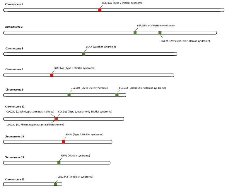
The Stickler syndromes are a group of genetic connective tissue disorders associated with an increased risk of rhegmatogenous retinal detachment, deafness, cleft palate, and premature arthritis. This review article focuses on the molecular genetics of the autosomal dominant forms of the disease. Pathogenic variants in COL2A1 causing Stickler syndrome usually result in haploinsufficiency of the protein, whereas pathogenic variants of type XI collagen more usually exert dominant negative effects. The severity of the disease phenotype is thus dependent on the location and nature of the mutation, as well as the normal developmental role of the respective protein.
Keywords: COL11A1; COL2A1; Stickler syndrome; giant retinal tear; retinal detachment; vitreous.
Conflict of interest statement
The authors declare no conflict of interest.
Figures


Similar articles
-
Inherited and de novo biallelic pathogenic variants in COL11A1 result in type 2 Stickler syndrome with severe hearing loss.Mol Genet Genomic Med. 2020 Sep;8(9):e1354. doi: 10.1002/mgg3.1354. Epub 2020 Jun 24. Mol Genet Genomic Med. 2020. PMID: 32578940 Free PMC article.
-
Autosomal Recessive Stickler Syndrome.Genes (Basel). 2022 Jun 24;13(7):1135. doi: 10.3390/genes13071135. Genes (Basel). 2022. PMID: 35885918 Free PMC article. Review.
-
Hearing Loss in Stickler Syndrome: An Update.Genes (Basel). 2022 Sep 1;13(9):1571. doi: 10.3390/genes13091571. Genes (Basel). 2022. PMID: 36140739 Free PMC article. Review.
-
Prevention of Blindness in Stickler Syndrome.Genes (Basel). 2022 Jun 26;13(7):1150. doi: 10.3390/genes13071150. Genes (Basel). 2022. PMID: 35885933 Free PMC article. Review.
-
Mutation survey and genotype-phenotype analysis of COL2A1 and COL11A1 genes in 16 Chinese patients with Stickler syndrome.Mol Vis. 2016 Jun 23;22:697-704. eCollection 2016. Mol Vis. 2016. PMID: 27390512 Free PMC article.
Cited by
-
Genetic and clinical profile of high myopia patients with rhegmatogenous retinal detachment.Front Genet. 2025 Apr 9;16:1485874. doi: 10.3389/fgene.2025.1485874. eCollection 2025. Front Genet. 2025. PMID: 40270540 Free PMC article.
-
Characteristics of a Three-Generation Family with Stickler Syndrome Type I Carrying Two Different COL2A1 Mutations.Genes (Basel). 2023 Mar 31;14(4):847. doi: 10.3390/genes14040847. Genes (Basel). 2023. PMID: 37107605 Free PMC article.
-
Unraveling the genetic collagen connection: clinical and therapeutic insights on genetic connective tissue disorders.Adv Rheumatol. 2024 Apr 25;64(1):32. doi: 10.1186/s42358-024-00373-z. Adv Rheumatol. 2024. PMID: 38664779 Review.
-
Glycosylation Modulates the Structure and Functions of Collagen: A Review.Molecules. 2024 Mar 22;29(7):1417. doi: 10.3390/molecules29071417. Molecules. 2024. PMID: 38611696 Free PMC article. Review.
-
From structure to therapy: the critical influence of cartilaginous endplates and microvascular network on intervertebral disc degeneration.Front Bioeng Biotechnol. 2024 Oct 28;12:1489420. doi: 10.3389/fbioe.2024.1489420. eCollection 2024. Front Bioeng Biotechnol. 2024. PMID: 39530056 Free PMC article. Review.
References
-
- Robin N.H., Moran R.T., Ala-Kokko L. GeneReviews(®) University of Washington; Washington, DC, USA: 2021. Stickler Syndrome.
-
- Stickler G.B., Belau P.G., Farrell F.J., Jones J.D., Pugh D.G., Steinberg A.G., Ward L.E. Hereditary Progressive Arthro-Ophthalmopathy. Mayo Clin. Proc. 1965;40:433–455. - PubMed
-
- Ahmad N.N., Ala-Kokko L., Knowlton R.G., Jimenez S.A., Weaver E.J., Maguire J.I., Tasman W., Prockop D.J. Stop codon in the procollagen II gene (COL2A1) in a family with the Stickler syndrome (arthro-ophthalmopathy) Proc. Natl. Acad. Sci. USA. 1991;88:6624–6627. doi: 10.1073/pnas.88.15.6624. - DOI - PMC - PubMed
Publication types
MeSH terms
Supplementary concepts
LinkOut - more resources
Full Text Sources
Medical
Miscellaneous
