Review
. 2022 Jun 7;23(12):6373.
doi: 10.3390/ijms23126373.

Structural and Molecular Kinetic Features of Activities of DNA Polymerases

Affiliations
Review

Structural and Molecular Kinetic Features of Activities of DNA Polymerases

Aleksandra A Kuznetsova et al. Int J Mol Sci. .

Abstract

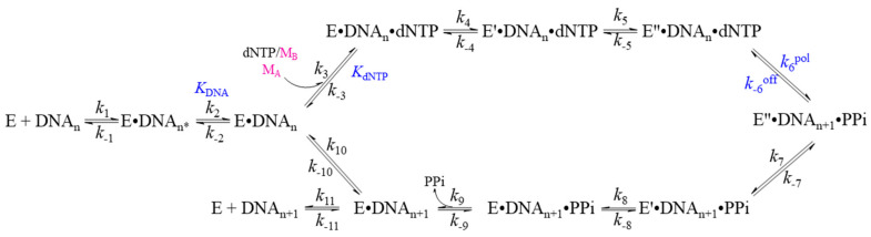
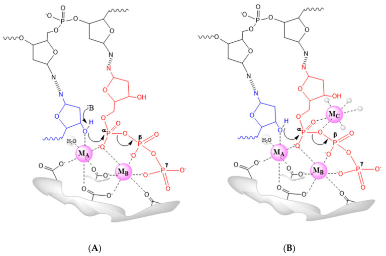
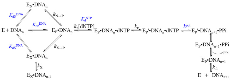
DNA polymerases catalyze DNA synthesis during the replication, repair, and recombination of DNA. Based on phylogenetic analysis and primary protein sequences, DNA polymerases have been categorized into seven families: A, B, C, D, X, Y, and RT. This review presents generalized data on the catalytic mechanism of action of DNA polymerases. The structural features of different DNA polymerase families are described in detail. The discussion highlights the kinetics and conformational dynamics of DNA polymerases from all known polymerase families during DNA synthesis.

Keywords: DNA polymerase; catalytic mechanism; kinetics; protein–DNA interaction; structural family.


Conflict of interest statement

The authors declare no conflict of interest.

Figures

Scheme 1
Scheme 1
The kinetic mechanism of dNTP incorporation into a DNA primer by polymerase.
Figure 1
Figure 1
The proposed two-metal-ion mechanism (A) and three-metal-ion mechanism (B). The incoming nucleotide is colored red, template and primer DNA are colored black and blue, respectively, enzyme moieties are colored grey, metal ions are magenta.
Figure 2
Figure 2
Three-dimensional structure of DNA polymerase I from E. coli: (A) holo enzyme (PDB ID 1ktq); (B) binary Pol-DNA (PDB ID 4ktq); (C) ternary Pol-DNA-dNTP (PDB ID 1qtm) complexes. The “thumb” and “fingers” subdomains are colored green and blue, respectively. The “palm” subdomain is red, “exonuclease” subdomain is grey, and DNA is magenta; (D,E) a detailed comparison of the active-site regions in the binary and ternary complexes. In each case, the protein is shown in beige, with the O-helix in purple. The incoming dNTP is colored in brown.
Figure 3
Figure 3
Three-dimensional structure of the binary complex of (A) Pol γ (PDB ID 5c52), (B) polymerase domain of Pol θ (PDB ID 4x0p), (C) Pol ν (PDB ID 4xvm) with DNA. The thumb (green), palm (red), fingers (blue), and exonuclease (gray) domains are presented. For Pol γ the spacer domain (orange) presents a unique structure. Structure of the heterotrimeric Pol γ holoenzyme contains one catalytic subunit and the Pol γB dimers (proximal (pink) and distal (light green) monomers).
Figure 4
Figure 4
Three-dimensional structure of (A) the holo Pol α with primase (PDB ID 5exr), and (B) binary complex of catalytic subunit p180core with DNA–RNA duplex (PDB ID 4q5v). Pol α is composed of two polypeptides: the catalytic subunit (p180) and the accessory B-subunit (p70). p180 contains two domains, the catalytic (p180core) and the C-terminal (CTD, light green) domains. CTD connects the catalytic domain with B-subunit and primase, and contains two conserved zinc-binding modules. The p180core consists of five subdomains: N-terminal (orange), thumb (green), palm (red), fingers (blue) and exonuclease-like (gray) domains. DNA–RNA duplex is dark magenta. The N terminus of p180 is predicted to be poorly folded and does not participate in primer synthesis. Human primase consists of catalytic (p49, purple) and regulatory (p58, pink) subunits.
Scheme 2
Scheme 2
The kinetic mechanism of incorporation of the correct nucleotide as catalyzed by the holoenzyme of human DNA polymerase ε [71].
Figure 5
Figure 5
The cryo-EM structure of (A) the E. coli replicative DNA polymerase complex (DNA polymerase III alpha, beta, epsilon, tau) (PDB ID 5fku), and (B) the crystal structure of the alpha-subunit–DNA complex (PDB ID 3e0d). The holoenzyme is composed of 10 subunits (α, β, ε, θ, δ′, γ, τ, χ, and ψ), together with the helicase DnaB and the RNA primase DnaG form the replisome. Pol IIIα contains six domains with different functions: the N-terminal Zn2+-dependent 3′-5′ co-proofreading exonuclease polymerase and histidinol phosphatase (PHP) domain (gray); the catalytic palm domain (red); the incoming nucleotide-interacting fingers domain (blue); the nascent DNA gripping-thumb domain (green); the β-sliding clamp-binding domain (orange); and the CTD (cyan), which contains an oligonucleotide-binding OB fold and a possible external clamp-binding site at the extreme C terminus (cyan).
Scheme 3
Scheme 3
Minimal single-nucleotide incorporation reaction pathway for PolC (Sau-PolC-∆N∆Exo) from Staphylococcus aureus [79].
Figure 6
Figure 6
The cryo-EM structure of the DNA-bound Pol D-PCNA processive complex (PDB ID 6t8h). PCNA trimer is colored blue. Overview of Pol D domain organization: DP1 includes OB (orange) and PDE (yellow); DP2 includes DPBB-1 and DPBB-2 (black), clamp-1 and clamp-2 domains (cyan), KH-like (green)—which is connected to the Anchor domain (dark green)—and Accessory-1 and Accessory-2 domains (grey).
Scheme 4
Scheme 4
Minimal single-nucleotide incorporation reaction pathway for Pol β and Pol λ.
Figure 7
Figure 7
Three-dimensional structure of the binary complex of (A) Pol β (PDB ID 1bpy), (B) Pol λ (PDB ID 1xsn), (C) Pol µ(PDB ID 5tyw), and (D) TdT (PDB ID 4i2a) with DNA. The thumb (green), palm (red), fingers (blue) and exonuclease (gray) domains are presented.
Figure 8
Figure 8
Three-dimensional structure of the complex of (A) Dpo4 (PDB ID 2agp), (B) Dbh (PDB ID 3bq1), (C) Pol κ (PDB ID 3pzp), (D) Pol η (PDB ID 4ed8), and (E) Pol ι (PDB ID 3gv8) with DNA. The thumb (green), palm (red), and fingers (blue) domains are presented; for Pol κ, the unique N-clasp domain at the N-terminus is presented (yellow).
Figure 9
Figure 9
Three-dimensional structure of HIV-1 reverse transcriptase (RT) heterodimer in complex with a double stranded RNA (PDB ID 6hak). The thumb (green), palm (red), fingers (blue), RNase H (grey), and connection (yellow) domains are presented.
Scheme 5
Scheme 5
Minimal single-nucleotide incorporation reaction pathway for HIVRT.

References

    1. Steitz T.A. DNA Polymerases: Structural Diversity and Common Mechanisms. J. Biol. Chem. 1999;274:17395–17398. doi: 10.1074/jbc.274.25.17395. - DOI - PubMed
    1. Raper A.T., Reed A.J., Suo Z. Kinetic Mechanism of DNA Polymerases: Contributions of Conformational Dynamics and a Third Divalent Metal Ion. Chem. Rev. 2018;118:6000–6025. doi: 10.1021/acs.chemrev.7b00685. - DOI - PubMed
    1. Berdis A.J. Mechanisms of DNA Polymerases. Chem. Rev. 2009;109:2862–2879. doi: 10.1021/cr800530b. - DOI - PubMed
    1. Rothwell P.J., Waksman G. Structure and Mechanism of DNA Polymerases. Adv. Protein Chem. 2005;71:401–440. doi: 10.1016/S0065-3233(04)71011-6. - DOI - PubMed
    1. Steitz T.A. DNA- and RNA-Dependent DNA Polymerases. Curr. Opin. Struct. Biol. 1993;3:31–38. doi: 10.1016/0959-440X(93)90198-T. - DOI

LinkOut - more resources