. 2022 Jun 7;23(12):6382.
doi: 10.3390/ijms23126382.

Canine Circovirus Suppresses the Type I Interferon Response and Protein Expression but Promotes CPV-2 Replication

Affiliations

Canine Circovirus Suppresses the Type I Interferon Response and Protein Expression but Promotes CPV-2 Replication

Xiangqi Hao et al. Int J Mol Sci. .

Abstract

Canine circovirus (CanineCV) is an emerging virus in canines. Since the first strain of CanineCV was reported in 2012, CanineCV infection has shown a trend toward becoming a global epidemic. CanineCV infection often occurs with coinfection with other pathogens that may aggravate the symptoms of disease in affected dogs. Currently, CanineCV has not been successfully isolated by laboratories, resulting in a lack of clarity regarding its physicochemical properties, replication process, and pathogenic characteristics. To address this knowledge gap, the following results were obtained in this study. First, a CanineCV strain was rescued in F81 cells using infectious clone plasmids. Second, the Rep protein produced by the viral packaging rescue process was found to be associated with cytopathic effects. Additionally, the Rep protein and CanineCV inhibited the activation of the type I interferon (IFN-I) promoter, blocking subsequent expression of interferon-stimulated genes (ISGs). Furthermore, Rep was found to broadly inhibit host protein expression. We speculate that in CanineCV and canine parvovirus type 2 (CPV-2) coinfection cases, CanineCV promotes CPV-2 replication by inducing immunosuppression, which may increase the severity of clinical symptoms.

Keywords: canine circovirus; canine parvovirus; coinfection; interferon response; protein expression inhibition.


Conflict of interest statement

The authors declare no conflict of interest.

Figures

Figure 3
Figure 3
CanineCV and Rep inhibit IFN-I and ISG expression. F81 cells were transfected with different plasmids, total RNA was extracted from the cells at the indicated time points, and the mRNA levels of IFN-α, IFN-β (A,E), MxA and ISG15 (B,F) were measured by qPCR. In the dual luciferase assay, F81 cells were transfected with different plasmids for 24 h, inoculated with SeV and incubated for another 12 h, and lysed for determination of luciferase activity (C,D,GI). CanineCV and the Rep protein inhibited SeV-activated IFN-α, IFN-β, MxA, and ISG15 expression at the mRNA level. Second, the results of the dual luciferase assay showed that CanineCV and the Rep protein blocked the activation of the IFN-β and ISRE promoters. Finally, the Rep protein blocked the interferon signaling pathway through the IRF3 and NFκB pathways. * p < 0.05, ** p < 0.01, *** p < 0.001, **** p < 0.0001 compared with the empty vector transfection group.
Figure 4
Figure 4
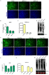
CanineCV and Rep inhibited protein expression. (A,E) F81 cells were cotransfected with EGFP-tagged protein expression plasmids and different viral plasmids. After 48 h, the cells were imaged under a fluorescence microscope (Exposure [ms] = 200; Gain = 1.5), and the fluorescence intensity, indicating the level of protein expression, was calculated using ImageJ (B,F). The results suggest that CanineCV and the Rep protein affected the expression of EGFP. (C,G) F81 cells were cotransfected with the pRL-TK plasmid and different viral plasmids. After 48 h, the cells were collected and Renilla luciferase activity was measured. Luciferase activity indirectly reflects the level of protein expression in cells. (D,H) Forty-eight hours after transfection, puromycin (10 μg/mL) was added to the medium and incubated at 37 °C for 30 min. The cells were washed twice with PBS and then lysed for WB analysis. Nascent peptides labeled with puromycin were detected with an anti-puromycin monoclonal antibody. An anti-Flag antibody was used to confirm viral protein expression (red), and an anti-GAPDH antibody was used as the internal reference. The WB analysis results showed that in the CanineCV rescue group and the Rep protein expression group, fewer peptides were labeled by puromycin, and cellular translation levels were low. * p < 0.05, *** p < 0.001, **** p < 0.0001.
Figure 1
Figure 1
CanineCV rescue. (A) Schematic diagram of the steps for constructing the CanineCV single gene copy and double gene copy infectious clone plasmids. pClone007-P1 and pClone007-P2 were constructed as intermediate plasmids. Eventually, pBSK-C1 and pBSK-C2 were used for virus rescue. (B) TEM images of F81 cells transfected with pBSK-C1 and pBSK-C2. F81 cells were inoculated into 6-well plates, and 2 µg of plasmid was transfected into the cells. Seventy-two hours after transfection, cells were collected and processed for observation. CanineCV particles are labeled with black arrows. Inclusion bodies are labeled using white arrows. TEM showed that pBSK-C2-transfected cells exhibited greater viral packaging. N, cell nucleus. The scale bar represents 500 nm. (C) The pBlueScript II SK (+) vector (2 µg) was digested according to the instructions, and electrophoresis was then performed to demonstrate the proper activity of DNase I. (D) qPCR was used to determine the number of rescued CanineCV particles at different time points, and both experimental groups showed the trend of an initial increase followed by a decrease in the number of virions.
Figure 2
Figure 2
CanineCV and Rep affected cell viability. (A) HEK-293T cells were transfected with empty vector, pBSK-C1, or pBSK-C2; CCK-8 reagent was then added at 24 h, 48 h, 72 h, and 96 h; and the OD450 value was measured after incubation. The production of CanineCV consistently affected cell viability. (B) HEK-293T cells were transfected with different viral expression plasmids. The growth status of each group of cells was directly imaged under an optical microscope, and Rep-protein-expressing cells showed CPEs. (C) A CCK-8 assay was performed to detect the effect of each viral protein on cell viability at 48 h. The cell viability in the Rep protein expression group was significantly lower than that in the other groups. The data shown in the figure are presented as the mean ± SD of three independent experiments. * p < 0.05, *** p < 0.001, **** p < 0.0001 and ns means no significance compared with the empty vector transfection group.
Figure 5
Figure 5
CanineCV and Rep promoted CPV-2 replication. F81 cells were transfected with the pBSK, pBSK-C1, or pBSK-C2 plasmid. Twenty-four hours later, the cells were inoculated with CPV-2 at an MOI of 1. After 24 h of infection, the CanineCV rescue group developed severe CPEs (A). Then, cells were harvested 48 h after viral infection, DNA was extracted, and qPCR was used to determine the CPV-2 copy number. The results suggested that the CPV-2 copy number in the CanineCV group was 2–3-fold higher than that in the control group (D). F81 cells were transfected with empty vector or the Rep plasmid. Twenty-four hours later, the cells were inoculated with CPV-2 at an MOI of 1. The Rep protein expression group showed significant cell lesions (B,C) and higher levels (2–3-fold) of CPV-2 replication (E,F). The data shown in the figure are presented as the mean ± SD of three independent experiments. **** p < 0.0001 compared with the empty vector transfection group.
Figure 6
Figure 6
Schematic diagram of the effect of CanineCV on the IFN-I signaling pathway and CPV-2 replication. Infectious clones were transfected into cells, after which viral particles were packaged depending on the characteristics of the rolling loop replication. Then, viral protein expression was initiated, the host cell IFN-I immune pathway was blocked, protein synthesis was reduced, and antiviral defenses were dysregulated. CPV-2 replicates at higher levels in intracellular environments whereas antiviral genes are expressed at low levels.

Similar articles

Cited by

References

    1. Kapoor A., Dubovi E.J., Henriquez-Rivera J.A., Lipkin W.I. Complete Genome Sequence of the First Canine Circovirus. J. Virol. 2012;86:7018. doi: 10.1128/JVI.00791-12. - DOI - PMC - PubMed
    1. Decaro N., Martella V., Desario C., Lanave G., Circella E., Cavalli A., Elia G., Camero M., Buonavoglia C. Genomic characterization of a circovirus associated with fatal hemorrhagic enteritis in dog, Italy. PLoS ONE. 2014;9:e105909. doi: 10.1371/journal.pone.0105909. - DOI - PMC - PubMed
    1. Li L., McGraw S., Zhu K., Leutenegger C.M., Marks S.L., Kubiski S., Gaffney P., Dela Cruz F.N., Jr., Wang C., Delwart E., et al. Circovirus in Tissues of Dogs with Vasculitis and Hemorrhage. Emerg. Infect. Dis. 2013;19:534–541. doi: 10.3201/eid1904.121390. - DOI - PMC - PubMed
    1. Van Kruiningen H.J., Heishima M., Kerr K.M., Garmendia A.E., Helal Z., Smyth J.A. Canine circoviral hemorrhagic enteritis in a dog in Connecticut. J. Vet. Diagn. Investig. 2019;31:732–736. doi: 10.1177/1040638719863102. - DOI - PMC - PubMed
    1. De Arcangeli S., Balboni A., Kaehler E., Urbani L., Verin R., Battilani M. Genomic Characterization of Canine Circovirus Detected in Red Foxes (Vulpes vulpes) from Italy using a New Real-Time PCR Assay. J. Wildl. Dis. 2020;56:239–242. doi: 10.7589/2018-11-270. - DOI - PubMed

Substances

LinkOut - more resources