. 2022 Jun 8;23(12):6409.
doi: 10.3390/ijms23126409.

The RelB-BLNK Axis Determines Cellular Response to a Novel Redox-Active Agent Betamethasone during Radiation Therapy in Prostate Cancer

Affiliations

The RelB-BLNK Axis Determines Cellular Response to a Novel Redox-Active Agent Betamethasone during Radiation Therapy in Prostate Cancer

Luksana Chaiswing et al. Int J Mol Sci. .

Abstract

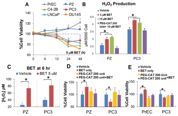
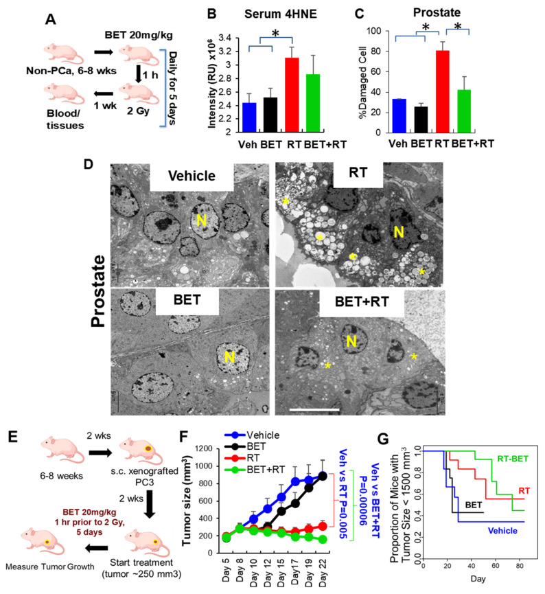
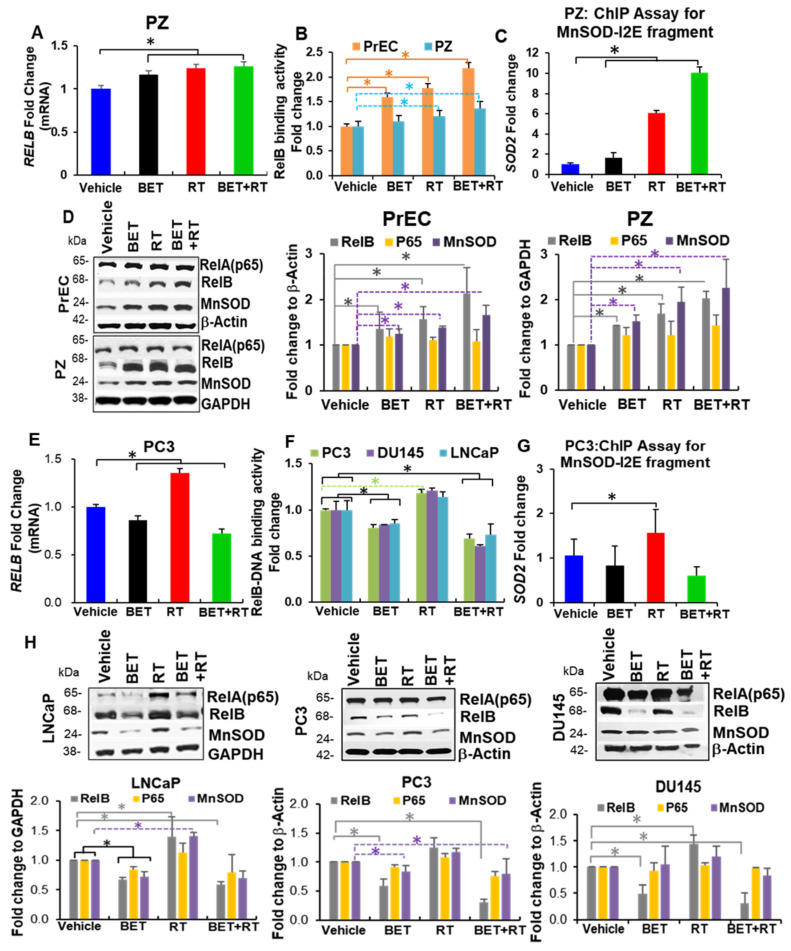
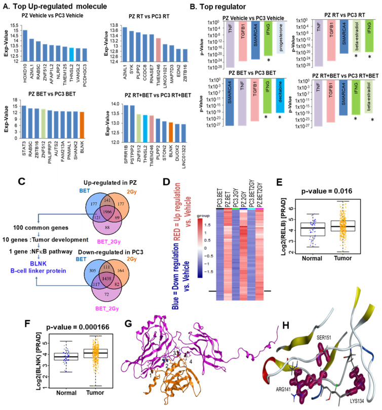
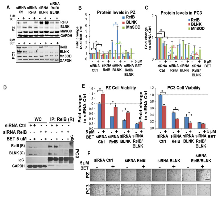
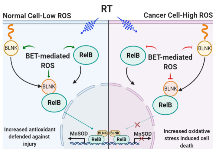
Aberrant levels of reactive oxygen species (ROS) are potential mechanisms that contribute to both cancer therapy efficacy and the side effects of cancer treatment. Upregulation of the non-canonical redox-sensitive NF-kB family member, RelB, confers radioresistance in prostate cancer (PCa). We screened FDA-approved compounds and identified betamethasone (BET) as a drug that increases hydrogen peroxide levels in vitro and protects non-PCa tissues/cells while also enhancing radiation killing of PCa tissues/cells, both in vitro and in vivo. Significantly, BET increases ROS levels and exerts different effects on RelB expression in normal cells and PCa cells. BET induces protein expression of RelB and RelB target genes, including the primary antioxidant enzyme, manganese superoxide dismutase (MnSOD), in normal cells, while it suppresses protein expression of RelB and MnSOD in LNCaP cells and PC3 cells. RNA sequencing analysis identifies B-cell linker protein (BLNK) as a novel RelB complementary partner that BET differentially regulates in normal cells and PCa cells. RelB and BLNK are upregulated and correlate with the aggressiveness of PCa in human samples. The RelB-BLNK axis translocates to the nuclear compartment to activate MnSOD protein expression. BET promotes the RelB-BLNK axis in normal cells but suppresses the RelB-BLNK axis in PCa cells. Targeted disruptions of RelB-BLNK expressions mitigate the radioprotective effect of BET on normal cells and the radiosensitizing effect of BET on PCa cells. Our study identified a novel RelB complementary partner and reveals a complex redox-mediated mechanism showing that the RelB-BLNK axis, at least in part, triggers differential responses to the redox-active agent BET by stimulating adaptive responses in normal cells but pushing PCa cells into oxidative stress overload.

Keywords: BLNK; RelB; betamethasone; radiation; redox state.


Conflict of interest statement

The authors declare no conflict of interest.

Figures

Figure 1
Figure 1
Library screening of FDA-approved drugs for compounds that induce PCa cell death while increasing non-cancer prostate cell viability through mediation of H2O2 production. PC3 cells and PZ cells were treated with library FDA-approved drugs for 24 h. (A) Cell viability. (B) Extracellular H2O2 based on Amplex Red assay. (C) Cell viability after drugs and radiation (4 Gy) treatment. Duplicate wells were tested per compound. (D) Drug candidates were chosen from a 786-compound library based on H2O2 production and the ability to exert opposite cytotoxic effects on non-cancer and cancer cells. One dot represents one compound. Please see supplementary excel file for details of each compound.
Figure 2
Figure 2
BET induces PCa cell death while increasing non-cancer prostate cell viability through mediation of H2O2 production. Cells were treated with 5 µM BET at various time points. (A) Cell viability based on MTT assay. (B) Extracellular H2O2 production (µM) based on Amplex Red method (24 h). (C) Intracellular H2O2 production was measured using the aminotriazole-(3-AT)-mediated inactivation of CAT method (6 h). (D,E) Cell viability after treatment with PEG-CAT prior to BET. * p-value ≤ 0.05 when compared with vehicle. n = 3.
Figure 3
Figure 3
Repurposing BET as a radioprotector for normal tissues while enhancing radiation efficacy for PCa in vivo. (A) Non-cancer nude mice were I.P. with BET alone or RT (2 Gy) 1 h prior, daily, for 5 days. Prostate, rectum, and blood were collected 1 week after treatment was completed. n = 5/group. (B) 4HNE protein adducts in serum. RU = Relative arbitrary unit. (C) Ultrastructural analysis of damaged prostate. (D) Representative photographs of damaged prostate. n = Nucleus. Star = Damaged mitochondria as indicated with loss of cristae and vacuolization of mitochondria. Bar = 6 µm. (E) PC3 xenograft tumor mice were I.P. with BET alone or RT (2 Gy) 1 h prior, daily, for 5 days. n = 10/group. (F) PC3 tumor size was measured every 2–3 days. (G) Survival fraction of PC3 xenograft tumor mice. * p-value ≤ 0.05 when compared to vehicle.
Figure 4
Figure 4
RelB expression determines radioprotector effect of BET in non-PCa cells and radio-killing effect of BET. PZ cells were treated with BET and/or RT (2 Gy) for 24 h. NF-kB transcription factor family expression levels and function were measured. (A) RT-PCR of RelB. (B) RelB binding activity. (C) Chip assay with RelB antibody to I2E promoter of MnSOD. (D) Representative Western blots and quantitative analysis of protein expression for PrEC cells and PZ cells. * p-value ≤ 0.05 when compared with vehicle. PCa cells (LNCaP, PC3, DU145) were treated with either BET and/or RT (2 Gy) for 24 h. NF-kB transcription factor family expression levels and function were measured. (E) RT-PCR of RelB of PC3 cells. (F) RelB binding activity of PCa cells. (G) Chip assay with RelB antibody to I2E promoter of MnSOD of PC3 cells. (H) Representative Western blots and quantitative analysis of protein expression of PCa cells. n = 3. * p-value ≤ 0.05 when compared with vehicle.
Figure 5
Figure 5
RNA sequencing identifies BLNK as a novel complementary protein that is upregulated by BET-mediated RelB expression. (A) Upregulated molecules that were most significantly changed (threshold exp-value 10). The rank order is determined by the indicated p-values. (B) 5 regulator pathways that were most significantly changed (threshold p-value 1.3). All these top regulators contain NF-kB family, and regulator star (*) contains BLNK. (C) Venn diagrams identified BLNK as the one of the proteins that is upregulated in PZ cells and downregulated in PC3 cells. (D) Heat map demonstrated ~2000 common genes that are diversely expressed in PC3 and PZ cells by all the treatments. Red = Upregulation vs. vehicle; Blue = Downregulation vs. vehicle, in PZ vs. PC3 cells. (E) RelB and (F) BLNK in PCa (n = 499) vs. normal prostate (n = 52) from TCGA database vs. vehicle. PARD = Prostate Adenocarcinoma. RELB and BLNK expressions are presented in log2-transformed transcripts per kilobase million (TPM) values. p-value calculated from linear mixed model. (G) PPI between RELB (magenta) and BLNK (orange). Contacts (1–4) given following: (1) Hydrogen bond at Cys 343 (BLNK) and Leu127 (RelB); (2) Ionic bond at Arg427 (BLNK) and Glu290 (RelB); (3) Hydrogen bond at His431 (BLNK) and Arg136 (RelB); and (4) Hydrogen bond at Arg448 (BLNK) and Cys389 (RelB). (H) Docking and binding analysis with ZDOCK and PyMol suggesting the interaction at the Y300 residue of RelB and the conserved R32 and R51 residues of BLNK.
Figure 6
Figure 6
RelB and BLNK exert their roles reversely in non-PCa cells vs. PCa cells with BET treatment. Cells were treated with BET for 24 h and then harvested for analysis. (A) Western blots of BLNK, RelB, and MnSOD upon BET treatment. (B) IP with RelB antibody in whole cells nuclear fraction (NE) after treatment with BET. R = Anti-rabbit antibody. G = Anti-goat antibody. (C) Proximity Ligation assay. Red = Binding of RelB and BLNK. Blue = Nucleus. Inserts indicate the binding of RelB and BLNK in the nuclei of PZ cells (yellow) but not in the nuclei of PC3 cells (yellow). Bar = 50 μm. (D) Representation of double immunogold electron microscopy of cells and quantification of gold bead number in nuclei. PZ cells or PC3 cells were labeled with anti-rabbit RelB antibody (6 nm gold beads, arrow heads) and anti-goat BLNK antibody (10 nm gold beads, arrow). The gold beads were localized primarily in the nuclei of PZ cells after BET treatment but less in the nuclei of PC3 cells. * p-value ≤ 0.05 when compared with vehicle. Twenty nuclei were counted per group.
Figure 7
Figure 7
BLNK interaction with RelB is a signal for BET protection of RT-induced injury in normal tissues. Cells were transfected with siRNA (Santa Cruz biotechnology) for 48 h and then treated with BET for 24 h. Western blot analysis (A) and protein quantification of (B) PC3 and (C) PZ cells. n = 2. (D) IP analysis confirmed a decrease in RelB:BLNK axis with RelB knockdown. WC = Whole cell lysates. R = Anti-rabbit antibody. G = Anti-goat antibody. (E) Trypan Blue assay and (F) representative photograph indicates a decrease in cell viability with siRNA against RelB, BLNK, or RelB + BLNK with or without BET treatment. n = 3. * p-value < 0.05 vs. non-BET. Scale bar = 50 µm.
Figure 8
Figure 8
Schematic of how BET-mediated ROS production sensitizes PCa cells toward death caused by RT while protecting non-PCa cells against injury from RT off-target effects. Aberrant redox homeostasis of cancer cells enables redox-modifying agent BET to enhance radiation therapy efficacy by selective sensitization. In normal cells under physiologic conditions, cellular redox status is kept at a low oxidizing level. A shift in cell redox status toward an oxidizing condition, from BET + RT, will stimulate the expression of RelB-BLNK and translocation to the nucleus, which leads to upregulation of the antioxidant system, including MnSOD. The upregulation of MnSOD maintains redox status in normal cells and promotes cell survival. To the contrary, cancer cells are usually under high oxidizing conditions. A comparable shift in ROS levels modulated by BET + RT to an extreme oxidizing condition will cause cell death. Green arrows = activation; Red line = inhibition.

References

    1. Siegel R.L., Miller K.D., Fuchs H.E., Jemal A. Cancer statistics, 2022. CA Cancer J. Clin. 2022;72:7–33. doi: 10.3322/caac.21708. - DOI - PubMed
    1. Chaiswing L., St Clair W.H., St Clair D.K. Redox Paradox: A Novel Approach to Therapeutics-Resistant Cancer. Antioxid. Redox Signal. 2018;29:1237–1272. doi: 10.1089/ars.2017.7485. - DOI - PMC - PubMed
    1. Conklin K.A. Chemotherapy-associated oxidative stress: Impact on chemotherapeutic effectiveness. Integr. Cancer Ther. 2004;3:294–300. doi: 10.1177/1534735404270335. - DOI - PubMed
    1. Barrera G. Oxidative stress and lipid peroxidation products in cancer progression and therapy. ISRN Oncol. 2012;2012:137289. doi: 10.5402/2012/137289. - DOI - PMC - PubMed
    1. Wilson C., Gonzalez-Billault C. Regulation of cytoskeletal dynamics by redox signaling and oxidative stress: Implications for neuronal development and trafficking. Front. Cell Neurosci. 2015;9:381. doi: 10.3389/fncel.2015.00381. - DOI - PMC - PubMed

MeSH terms