. 2022 Jun 9;23(12):6470.
doi: 10.3390/ijms23126470.

Comprehensive Integrated Single-Cell Whole Transcriptome Analysis Revealed the p-EMT Tumor Cells-CAFs Communication in Oral Squamous Cell Carcinoma

Affiliations

Comprehensive Integrated Single-Cell Whole Transcriptome Analysis Revealed the p-EMT Tumor Cells-CAFs Communication in Oral Squamous Cell Carcinoma

Nam Cong-Nhat Huynh et al. Int J Mol Sci. .

Abstract

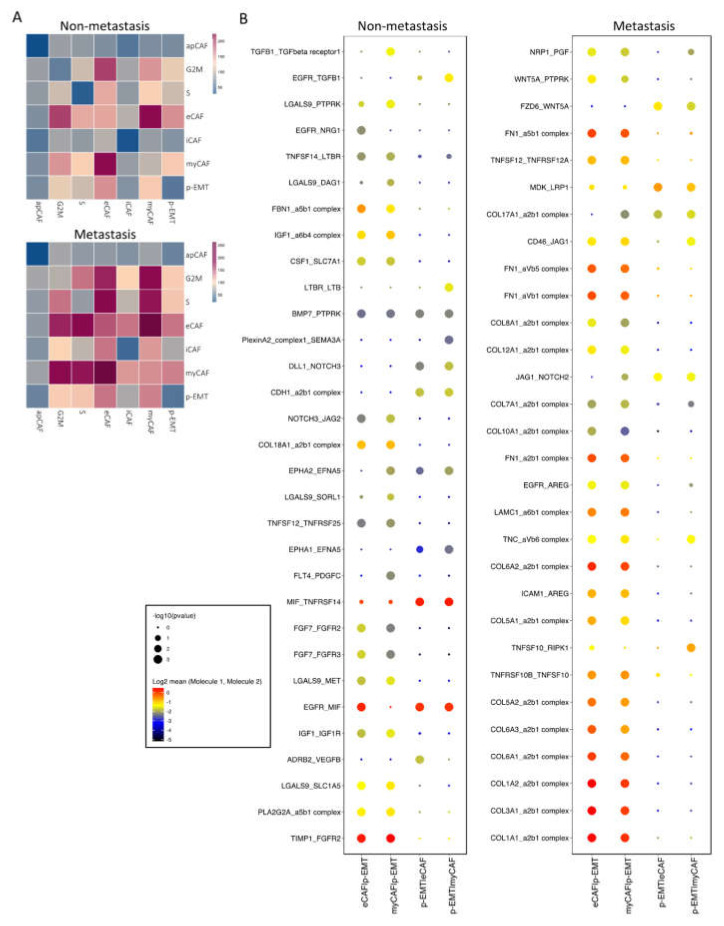
Cancer-associated fibroblasts (CAFs) and partial epithelial-mesenchymal transition (p-EMT) tumor cells are closed together and contribute to the tumor progression of oral squamous cell carcinoma (OSCC). In the present study, we deeply analyzed and integrated OSCC single-cell RNA sequencing datasets to define OSCC CAFs and p-EMT subpopulations. We highlighted the cell-cell interaction network of CAFs and p-EMT tumor cells and suggested biomarkers for the diagnosis and prognosis of OSCC during the metastasis condition. The analysis discovered four subtypes of CAFs: one p-EMT tumor cell population, and cycling tumor cells as well as TNFSF12-TNFRSF25/TNFRSF12A interactions between CAFs and p-EMT tumor cells during tumor metastasis. This suggests the prediction of therapeutically targetable checkpoint receptor-ligand interactions between CAFs and p-EMT tumor cells in OSCC regarding the metastasis status.

Keywords: cancer-associated fibroblasts; metastasis; oral squamous cell carcinoma; partial epithelial–mesenchymal transition; single-cell RNA sequencing.


Conflict of interest statement

The authors have no conflicts of interest relevant to this article.

Figures

Figure 1
Figure 1
Single-cell RNA-seq integration of oral squamous cell carcinoma data. (A) Integrated cells were visualized by UMAP with 15 main cell types assigned names based on canonical markers. (B) Integrated scRNA-seq data included 16,817 non-lymph node metastasis cells (0, N0) and 13,183 lymph node metastasis cells (1, N1 to Nx). (C) Dotplot of the average expression of the main markers of each cell type in oral squamous cell carcinoma integration data.
Figure 2
Figure 2
Sub-clusters of fibroblasts and epithelial cells. (A) Cells were sub-clustered into 7 sub-types and visualized by UMAP. (B,C) Integrated data included 2,270 non-lymph node metastasis cells (0, N0) and 3,239 lymph node metastasis cells (1, N1 to Nx). (D) Dotplot of average expression of the main markers of each sub-type in integration data. (E) Expressions of distinguished markers in non- (0) vs. metastasis (1) OSCC. (F) Differentiation of gene expression of eCAFs, myCAFs, and p-EMT tumor cells by pseudo bulk RNA-seq analysis in non- (X0) vs. metastasis (X1) conditions. Red dots are significantly different genes; black dots are total genes; blue labels are the top 20 significant different genes by adjusted p value.
Figure 3
Figure 3
Cell–cell communication analysis in sub-clusters of fibroblasts and epithelial cells. (A) Heatmap of all interactions in non- vs. metastasis conditions by CellPhoneDB analysis. (B) Top exclusive 30 significant interactions upregulated only in non- or metastasis conditions between p-EMT cluster and eCAF/myCAF clusters (dot’s sizes illustrate the -log10(pvalue), in which the bigger size presents the lower p value; dot’s colors illustrate the mean interaction number, in which the redder color presents the higher number of mean interactions between 2 clusters.
Figure 4
Figure 4
WNT and NOTCH signaling pathways increased in metastasis p-EMT cluster and eCAF/myCAF clusters. (A) Significant WNT signaling pathway communications upregulated in metastasis conditions between p-EMT cluster and eCAF/myCAF clusters (with compared non-metastasis condition). (B) Significant NOTCH signaling pathway communications upregulated in metastasis condition between p-EMT cluster and eCAF/myCAF clusters (with compared non-metastasis condition). (C) WNT and NOTCH signaling pathway-related gene expressions in integrated single-cell RNA-seq of fibroblasts and epithelial cells in non- vs. metastasis conditions.
Figure 5
Figure 5
Different roles of TNFRSF25 and TNFRSF12A in non- and metastasis conditions of oral cancer. (A) Significant TNF signaling pathway communications were altered in non- vs. metastasis conditions between p-EMT and eCAF/myCAF clusters. (B) TNF signaling pathway-related gene expression in integrated single-cell RNA-seq of fibroblasts and epithelial cells in non- vs. metastasis conditions. (C) Survival plots of head and neck cancer patients with high TNFRSF25 (left panel) and TNFRSF12A (right panel) expression from TCGA database. (D) Proportion of TNFRSF12A expression in head and neck cancer patients from Human Protein Atlas. (E) Top 100 TNFRSF12A-correlated genes in integrated single-cell RNA-seq of fibroblasts and epithelial cells. (F) Co-expression (left) and scatter (right) plots of TNFRSF12A (red) and SNAI2 (green) in integrated single-cell RNA-seq of fibroblasts and epithelial cells in non- (0) vs. metastasis (1) conditions. (G) Correlation plots of TNFRSF12A and SNAI2 gene expressions in HNSCC non-tumor samples (normal cells) and tumor tissues from TCGA database. (H) Conceptual diagram (visualized by BiorRender) of p-EMT cells and CAFs communication in non- vs. metastasis oral squamous cell carcinomas via TWEAK (TNFSF12)-DR3 (TNFRSF25)/TWEAKR (TNFRSF12A).

References

    1. Hsieh Y.-P., Wu Y.-H., Cheng S.-M., Lin F.-K., Hwang D.-Y., Jiang S.-S., Chen K.-C., Chen M.-Y., Chiang W.-F., Liu K.-J., et al. Single-Cell RNA Sequencing Analysis for Oncogenic Mechanisms Underlying Oral Squamous Cell Carcinoma Carcinogenesis with Candida albicans Infection. Int. J. Mol. Sci. 2022;23:4833. doi: 10.3390/ijms23094833. - DOI - PMC - PubMed
    1. Fox S.A., Vacher M., Farah C.S. Transcriptomic Biomarker Signatures for Discrimination of Oral Cancer Surgical Margins. Biomolecules. 2022;12:464. doi: 10.3390/biom12030464. - DOI - PMC - PubMed
    1. Bray F., Ferlay J., Soerjomataram I., Siegel R.L., Torre L.A., Jemal A. Global cancer statistics 2018: GLOBOCAN estimates of incidence and mortality worldwide for 36 cancers in 185 countries. CA Cancer J. Clin. 2018;68:394–424. doi: 10.3322/caac.21492. - DOI - PubMed
    1. Lesterhuis W.J., Bosco A., Millward M.J., Small M., Nowak A.K., Lake R.A. Dynamic versus static biomarkers in cancer immune checkpoint blockade: Unravelling complexity. Nat. Rev. Drug Discov. 2017;16:264–272. doi: 10.1038/nrd.2016.233. - DOI - PubMed
    1. Huang L.Y., Hsieh Y.P., Wang Y.Y., Hwang D.Y., Jiang S.S., Huang W.T., Chiang W.F., Liu K.J., Huang T.T. Single-Cell Analysis of Different Stages of Oral Cancer Carcinogenesis in a Mouse Model. Int. J. Mol. Sci. 2020;21:8171. doi: 10.3390/ijms21218171. - DOI - PMC - PubMed

MeSH terms