Review
. 2022 Jun 10;23(12):6489.
doi: 10.3390/ijms23126489.

EGFR-Mutated Non-Small Cell Lung Cancer and Resistance to Immunotherapy: Role of the Tumor Microenvironment

Affiliations
Review

EGFR-Mutated Non-Small Cell Lung Cancer and Resistance to Immunotherapy: Role of the Tumor Microenvironment

Clelia Madeddu et al. Int J Mol Sci. .

Abstract

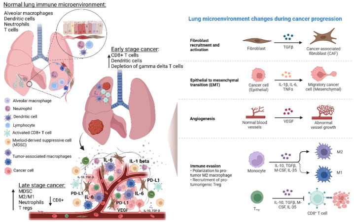
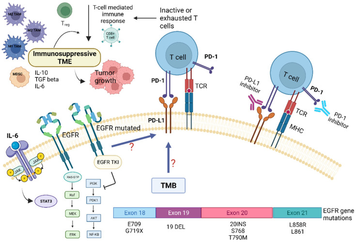
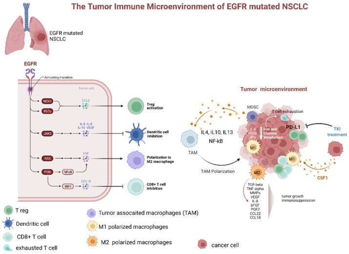
Lung cancer is a leading cause of cancer-related deaths worldwide. About 10-30% of patients with non-small cell lung cancer (NSCLC) harbor mutations of the EGFR gene. The Tumor Microenvironment (TME) of patients with NSCLC harboring EGFR mutations displays peculiar characteristics and may modulate the antitumor immune response. EGFR activation increases PD-L1 expression in tumor cells, inducing T cell apoptosis and immune escape. EGFR-Tyrosine Kinase Inhibitors (TKIs) strengthen MHC class I and II antigen presentation in response to IFN-γ, boost CD8+ T-cells levels and DCs, eliminate FOXP3+ Tregs, inhibit macrophage polarization into the M2 phenotype, and decrease PD-L1 expression in cancer cells. Thus, targeted therapy blocks specific signaling pathways, whereas immunotherapy stimulates the immune system to attack tumor cells evading immune surveillance. A combination of TKIs and immunotherapy may have suboptimal synergistic effects. However, data are controversial because activated EGFR signaling allows NSCLC cells to use multiple strategies to create an immunosuppressive TME, including recruitment of Tumor-Associated Macrophages and Tregs and the production of inhibitory cytokines and metabolites. Therefore, these mechanisms should be characterized and targeted by a combined pharmacological approach that also concerns disease stage, cancer-related inflammation with related systemic symptoms, and the general status of the patients to overcome the single-drug resistance development.

Keywords: EGFR mutations; immunotherapy resistance; non-small cell lung cancer; tumor microenvironment; tumor-associated macrophages; tyrosine kinase inhibitor.


Conflict of interest statement

The authors declare no conflict of interest.

Figures

Figure 1
Figure 1
Lung microenvironment changes from physiological state to NSCLC development and progression. Normal lung microenvironment includes epithelial cells, smooth muscle cells, fibroblast, endothelial cells, and immune cells such as dendritic cells, neutrophils, T cells, and alveolar macrophages. The latter contribute to maintain immunological homeostasis, but they can also promote inflammation, and thus, development of premalignant lung lesions and carcinogenesis. During NSCLC development, the tumor microenvironment (TME) changes, thus contributing to inflammation angiogenesis, immune modulation, and therefore promoting NSCLC progression, metastasis, and prognosis. In particular, the immune TME, through specific reprogramming and modulation of T cells, tumor-associated macrophages (TAMs) and myeloid cell populations exert crucial tumor-promoting or tumor-suppressing activities. The switch from tumor immune surveillance to cancer immune escape is characterized by the recruitment of regulatory T-cells (Tregs) and upregulation of MDSCs. In addition, TAMs (M1 and M2 polarized cells), and neutrophils play a key role in the mechanisms of immune escape. In particular, they contribute to create a proinflammatory TME that strongly affects the immune response efficiency. Abbreviations: MDSCs—myeloid-derived suppressor cells; IL—Interleukin; VEGF—vascular endothelial growth factor; PD-L—programmed death-ligand; TGF—tumor growth factor; CSF—colony-stimulating factor; M-CSF—macrophage-colony stimulating growth factor. Created with BioRender.com (https://biorender.com/, accessed on 17 May 2022).
Figure 2
Figure 2
Role of tumor microenvironment in EGFR-mutated NSCLC in influencing resistance pathways to targeted TKI treatment and potential targets for immunotherapy. EGFR mutations are associated with immunosuppressive TME, lower tumor-mutation burden (TMB), and increased PD-L1 expression. EGFR mutations may promote cancer immune escape through modulation of the PD-1/PD-L1 pathway, which in turn determine T-cells inactivity and/or exhaustion. This also leads to EGFR-TKI resistance. In addition, EGFR mutations influence several TME components, such as tumor-infiltrating lymphocytes (TILs), Tregs, MDSCs, TAMs, and immunoregulatory/proinflammatory cytokines, i.e., IL-6. The latter, through the activation of the STAT-3 intracellular pathway, contribute to tumor growth and resistance to targeted therapies. Abbreviations: AKT—serine-threonine kinase; EGFR, epidermal growth factor receptor; ERK—extracellular signal-regulated kinase; IL—Interleukin; JAK—Janus kinase; MHC—major histocompatibility complex; MEK—mitogen-activated protein kinase; MDSC—myeloid-derived suppressor cells; NF-kB, nuclear factor kappa B; PI3K—phosphatidylinositol-4,5-bisphosphate 3-kinase; PD-1—programmed death; PD-L1—programmed death ligand-1; TKI—Tyrosine kinase inhibitors; Treg—regulatory T-cell; STAT3—signal transducer and activator of transcription 3; TCR—T-cell receptor; TMB—tumor mutational burden. Created with BioRender.com (https://biorender.com/, accessed on 17 May 2022).
Figure 3
Figure 3
Dynamic changes of tumor microenvironment (TME) of EGFR-mutated NSCLC during tyrosine kinase inhibitor treatment. TME of EGFR mutated adenocarcinoma is typically characterized by prevalence of M2 polarized macrophage, low levels of CD8+ cells, increased number of Treg, and upregulation of PD-L1. The latter, especially if associated with macrophage-mediated inflammation particularly through IL-6 and increased ROS levels, contributes to T-cell exhaustion. Additionally, several factors secreted by M2 polarized TAMs (as TGFbeta, TNFalpha, MMPs, VEGF, IL-8, bFGF, PGE2, CCL22, and CCL18). These factors contribute to tumor progression and immunodepression. The TKI treatment has been associated with a decrease in PD-L1 expression, lowering of Treg, and promotion of TAM polarization from M2 to M1 phenotype. Abbreviations: EGFR—epidermal growth factor receptor; NSCLC—non-small cell lung cancer; JAK—Janus Kinase; PI3K—phosphatidylinositol-3 kinase; NF-kB—nuclear factor-κB; IRF1—interferon regulatory factor 1; IL—interleukin; TAM—tumor-associated macrophages; MDCS—myeloid-derived suppressor cells; PD-L1—programmed death-ligand 1; ROS—reactive oxygen species; IFN—interferon; TKI—tyrosine kinase inhibitor; CSF1—colony stimulating factor 1; TGF—tumor growth factor; TNF—tumor necrosis factor; MMP—matrix metalloproteases; VEGF—vascular endothelial growth factor; PGE—prostaglandin E; CCL—C-C-motif ligand. Created with BioRender.com (https://biorender.com/, accessed on 17 May 2022).

References

    1. Siegel R.L., Miller K.D., Fuchs H.E., Jemal A. Cancer Statistics, 2021. CA Cancer J. Clin. 2021;71:7–33. doi: 10.3322/caac.21654. - DOI - PubMed
    1. Mansuet-Lupo A., Alifano M., Pécuchet N., Biton J., Becht E., Goc J., Germain C., Ouakrim H., Régnard J.F., Cremer I., et al. Intratumoral immune cell densities are associated with lung adenocarcinoma gene alterations. Am. J. Respir. Crit. Care Med. 2016;194:1403–1412. doi: 10.1164/rccm.201510-2031OC. - DOI - PubMed
    1. Lynch T.J., Bell D.W., Sordella R., Gurubhagavatula S., Okimoto R.A., Brannigan B.W., Harris P.L., Haserlat S.M., Supko J.G., Haluska F.G., et al. Activating mutations in the epidermal growth factor receptor underlying responsiveness of non-small-cell lung cancer to gefitinib. N. Engl. J. Med. 2004;350:2129–2139. doi: 10.1056/NEJMoa040938. - DOI - PubMed
    1. Wang D.-H., Lee H.-S., Yoon D., Berry G., Wheeler T.M., Sugarbaker D.J., Kheradmand F., Engleman E., Burt B.M. Progression of EGFR-Mutant Lung Adenocarcinoma is Driven By Alveolar Macrophages. Clin. Cancer Res. 2017;23:778–788. doi: 10.1158/1078-0432.CCR-15-2597. - DOI - PubMed
    1. Ohashi K., Maruvka Y.E., Michor F., Pao W. Epidermal Growth Factor Receptor Tyrosine Kinase Inhibitor–Resistant Disease. J. Clin. Oncol. 2013;31:1070–1080. doi: 10.1200/JCO.2012.43.3912. - DOI - PMC - PubMed

MeSH terms