Review
. 2022 Jun 11;23(12):6539.
doi: 10.3390/ijms23126539.

TLR/WNT: A Novel Relationship in Immunomodulation of Lung Cancer

Affiliations
Review

TLR/WNT: A Novel Relationship in Immunomodulation of Lung Cancer

Aina Martín-Medina et al. Int J Mol Sci. .

Abstract

The most frequent cause of death by cancer worldwide is lung cancer, and the 5-year survival rate is still very poor for patients with advanced stage. Understanding the crosstalk between the signaling pathways that are involved in disease, especially in metastasis, is crucial to developing new targeted therapies. Toll-like receptors (TLRs) are master regulators of the immune responses, and their dysregulation in lung cancer is linked to immune escape and promotes tumor malignancy by facilitating angiogenesis and proliferation. On the other hand, over-activation of the WNT signaling pathway has been reported in lung cancer and is also associated with tumor metastasis via induction of Epithelial-to-mesenchymal-transition (EMT)-like processes. An interaction between both TLRs and the WNT pathway was discovered recently as it was found that the TLR pathway can be activated by WNT ligands in the tumor microenvironment; however, the implications of such interactions in the context of lung cancer have not been discussed yet. Here, we offer an overview of the interaction of TLR-WNT in the lung and its potential implications and role in the oncogenic process.

Keywords: TLR; WNT; lung cancer.


Conflict of interest statement

The authors declare no conflict of interest.

Figures
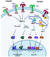
Figure 1
Figure 1
TLR2, TLR1, TLR4, TLR5, and TLR6 are expressed on the outer cell membrane; they recognize both extracellular pathogen-associated molecular patterns (PAMPs) and damage-associated molecular patterns (DAMPs). TLR3, TLR7, TLR8, and TLR9 are located in endosomal compartments where they bind to nucleic acids. All TLRs use the myeloid differentiation primary response protein 88 (MyD88) pathway, except TLR3, whose signaling depends on the IFN-α (TRIF) pathway that contains the TIR domain-containing adapter. TLR activates the MyD88-dependent canonical pathway that, through recruitment of TNF receptor-associated factors (TRAF) 6, leads to the activation of the transcription factor NF-KB, mitogen-activated protein kinase (MAPK), and activator protein-1 (AP-1), with the consequent induction of the production of proinflammatory cytokines. Intracellular TLRs are primarily involved in the type I interferon response. TLR7, TLR8, and TLR9 activate interferon regulatory factor 7 (IRF7) through the recruitment of TRAF6. TLR3 and TLR4 use the TRIF-dependent pathway. TRAF3 is activated and consequently activates IRF3, resulting in the induction of type I interferon (IFN) production. Created with BioRender.com (accessed on 24 February 2022).
Figure 2
Figure 2
(A): TLR activation on myeloid-derived suppressor cells (MDSCs), B regulatory cells (Breg), and T regulatory cells (Treg) leads to an immunosuppressive effect. Indeed, MDSCs can induce macrophages to release nitric oxide (NO) that suppresses the activity of T CD8+ lymphocytes. Moreover, activated MDSCs, Bregs, and Tregs release IL-6 and IL-10 with a consequent synergic inhibitory effect on T CD8+ cells. (B): TLR expressed on tumor cells can bind DAMPs/PAMPs and activate intracellular signaling, such as nuclear factor kappa-light-chain-enhancer of activated B cells (NF-kB), AP-1, Akt/PI3K, extracellular signal-regulated kinase (ERK)1/2, and MAPK. The consequence is a pro-tumor effect due to the release of IL-6, IL-8, and VEGF and the promotion of proliferation, angiogenesis, and protection from apoptosis. Continuous arrows indicate activation or secretion. Created with BioRender.com (accessed on 4 May 2022).
Figure 3
Figure 3
Scheme of the three WNT pathways. Created by BioRender.com and modified from Koni et al. [66] and Hiremath et al. [67].
Figure 4
Figure 4
Interaction between tumor-derived WNT and immune cells. Increased production of WNT by tumor cells (right part of the figure) stimulates an autocrine pathway that promotes the secretion of proinflammatory cytokines (IL-6) and chemokines (MCP-1). Both cytokines and WNT are maintained at a high level of expression by a self-feeding loop. Chemokines are critical for populating the tumor microenvironment (TME) with immune cells. The different cytokines induced by WNT, together with the WNT ligand itself, would stimulate other cells in the TME, such as tumor-associated fibroblasts or endothelial cells that further amplify the effects of WNT. At a later stage, once immune cells are recruited to the TME, WNT begins to play a different role (left part of the figure). In this new scenario, WNT activates a TLR/MyD88/p50 pathway, promoting the synthesis of the anti-inflammatory cytokine IL-10. As a result, WNT induces immunosuppression and the formation of tolerogenic mononuclear phagocytes. Created with BioRender.com (accessed on 21 March 2022).
Figure 5
Figure 5
Immunomodulatory effects of WNT on the tumor microenvironment. The autocrine effect of WNT (garnet arrow) drives the secretion of various cytokines and chemokines by tumor cells (blue arrows), which in turn promote different processes (in red) and chemotaxis of various cell types (orange arrow). WNT also promotes IL-10 secretion by dendritic cells (DC) and macrophages. WNT released by tumor cells promotes (black lines with arrowheads) or inhibits (black lines) maturation and/or differentiation (red arrows) of immune cells. The effects associated with WNT are angiogenesis, inflammation, and immunosuppression, as well as proliferation and survival of tumor cells. Created with BioRender.com (accessed on 21 March 2022).

Similar articles

Cited by

References

    1. Siegel R.L., Miller K.D., Jemal A. Cancer statistics, 2020. CA Cancer J. Clin. 2020;70:7–30. doi: 10.3322/caac.21590. - DOI - PubMed
    1. Lemjabbar-Alaoui H., Hassan O.U., Yang Y.-W., Buchanan P. Lung cancer: Biology and treatment options. Biochim. Biophys. Acta. 2015;1856:189–210. doi: 10.1016/j.bbcan.2015.08.002. - DOI - PMC - PubMed
    1. Herbst R.S., Morgensztern D., Boshoff C. The biology and management of non-small cell lung cancer. Nature. 2018;553:446–454. doi: 10.1038/nature25183. - DOI - PubMed
    1. Travis W.D. Update on small cell carcinoma and its differentiation from squamous cell carcinoma and other non-small cell carcinomas. Mod. Pathol. 2012;25:S18–S30. doi: 10.1038/modpathol.2011.150. - DOI - PubMed
    1. Li X., Jiang S., Tapping R.I. Toll-like receptor signaling in cell proliferation and survival. Cytokine. 2010;49:1–9. doi: 10.1016/j.cyto.2009.08.010. - DOI - PMC - PubMed

Substances