Case Reports
. 2022 Jun 13;23(12):6590.
doi: 10.3390/ijms23126590.

Large Benefit from Simple Things: High-Dose Vitamin A Improves RBP4-Related Retinal Dystrophy

Affiliations
Case Reports

Large Benefit from Simple Things: High-Dose Vitamin A Improves RBP4-Related Retinal Dystrophy

Vasily M Smirnov et al. Int J Mol Sci. .

Abstract

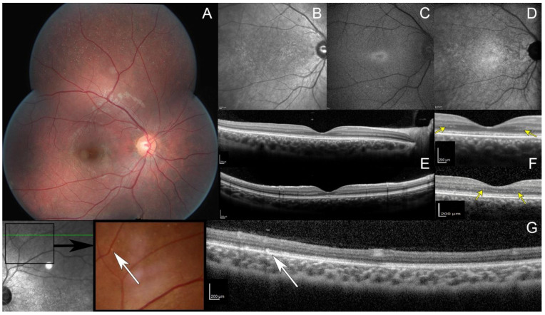
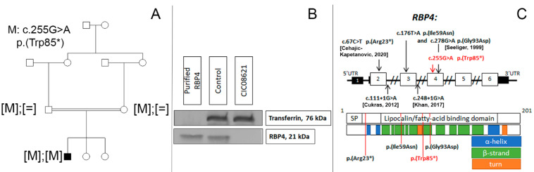
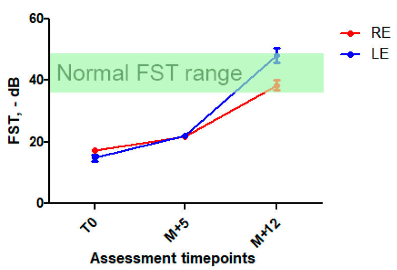
Inherited retinal diseases (IRD) are a group of heterogeneous disorders, most of which lead to blindness with limited therapeutic options. Pathogenic variants in RBP4, coding for a major blood carrier of retinol, retinol-binding protein 4, are responsible for a peculiar form of IRD. The aim of this study was to investigate if retinal function of an RBP4-related IRD patient can be improved by retinol administration. Our patient presented a peculiar white-dot retinopathy, reminiscent of vitamin A deficient retinopathy. Using a customized next generation sequencing (NGS) IRD panel we discovered a novel loss-of-function homozygous pathogenic variant in RBP4: c.255G >A, p.(Trp85*). Western blotting revealed the absence of RBP4 protein in the patient’s serum. Blood retinol levels were undetectable. The patient was put on a high-dose oral retinol regimen (50,000 UI twice a week). Subjective symptoms and retinal function markedly and sustainably improved at 5-months and 1-year follow-up. Here we show that this novel IRD case can be treated by oral retinol administration.

Keywords: RBP4; fundus albipunctatus; inherited retinal degeneration; retinol treatment; retinol-binding protein.


Conflict of interest statement

The authors declare no conflict of interest.

Figures

Figure 1
Figure 1
Multimodal retinal imaging. (A) Fundus photo, multiple white dots scattered over the midperipheral retina. Note the absence of intraretinal pigment migration and the lack of retinal vessel attenuation. (B) Infrared reflectance image, macular granularity. (C) Short-wavelength fundus autofluorescence imaging obtained with significant averaging due to the generalized reduced autofluorescence, small peri-foveal hyperautofluorescent ring with indistinct borders. (D) Near infrared fundus autofluorescence, small hypoautofluorescent dots. (E,F) spectral domain optic coherence tomography (SD-OCT, top: horizontal scan; and bottom: vertical scan), hypo reflective and fragmented ellipsoid zone with no interdigitation zone; preserved outer nuclear layer thickness with an unusual hyper reflective band on both sides of the fovea (yellow arrows). (G) OCT (scan passing through white dots, white arrows), hyperreflective dots above the retinal pigment epithelium with a focal interruption of the ellipsoid zone.
Figure 2
Figure 2
(A) Novel biallelic RBP4 variant co-segregating with the disease. (B) Western blot analysis showing the absence of RBP4 in peripheral blood of our patient (CIC08621) carrying the homozygous nonsense variant in RBP4, compared to an unaffected control and recombinant RPB4. Transferrin is used as serum loading control. (C) RBP4 gene and protein structure. SP—signal peptide domain. Disulfide bonds (22-178, 88-192, 138-147) are not shown. Previously reported and novel (in red) variants linked with inherited retinal degeneration [9,10].
Figure 3
Figure 3
Full stimulus threshold before and after high-dose vitamin A intake. Mean FST ± SD at first assessment, at M + 5 and M + 12 follow-up. A significant improvement in FST was observed at M + 5 (25 and 40% decrease) and was sustained at M + 12 (123 et 215% in RE and LE, respectively). In green, normal FST range [11,12].

Similar articles

Cited by

References

    1. Grant C.A., Berson E.L. Treatable Forms of Retinitis Pigmentosa Associated with Systemic Neurological Disorders. Int Ophthalmol. Clin. 2001;41:103–110. doi: 10.1097/00004397-200101000-00010. - DOI - PubMed
    1. Weleber R.G., Kurz D.E., Trzupek K.M. Treatment of Retinal and Choroidal Degenerations and Dystrophies: Current Status and Prospects for Gene-Based Therapy. Ophthalmol. Clin. N. Am. 2003;16:583–593. doi: 10.1016/S0896-1549(03)00072-5. - DOI - PubMed
    1. Chowers I., Banin E., Merin S., Cooper M., Granot E. Long-Term Assessment of Combined Vitamin A and E Treatment for the Prevention of Retinal Degeneration in Abetalipoproteinaemia and Hypobetalipoproteinaemia Patients. Eye. 2001;15:525–530. doi: 10.1038/eye.2001.167. - DOI - PubMed
    1. Balfoort B.M., Buijs M.J.N., Ten Asbroek A.L.M.A., Bergen A.A.B., Boon C.J.F., Ferreira E.A., Houtkooper R.H., Wagenmakers M.A.E.M., Wanders R.J.A., Waterham H.R., et al. A Review of Treatment Modalities in Gyrate Atrophy of the Choroid and Retina (GACR) Mol. Genet. Metab. 2021;134:96–116. doi: 10.1016/j.ymgme.2021.07.010. - DOI - PubMed
    1. Casalino G., Pierro L., Manitto M.P., Michaelides M., Bandello F. Resolution of Cystoid Macular Edema Following Arginine-Restricted Diet and Vitamin B6 Supplementation in a Case of Gyrate Atrophy. J. AAPOS. 2018;22:321–323. doi: 10.1016/j.jaapos.2017.12.016. - DOI - PubMed

Publication types

MeSH terms