

The State of the Art of Piezo1 Channels in Skeletal Muscle Regeneration
- PMID: 35743058
- PMCID: PMC9224226
- DOI: 10.3390/ijms23126616
The State of the Art of Piezo1 Channels in Skeletal Muscle Regeneration
Abstract
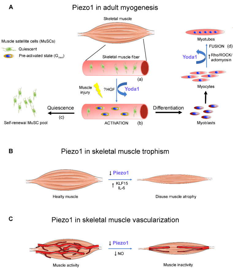
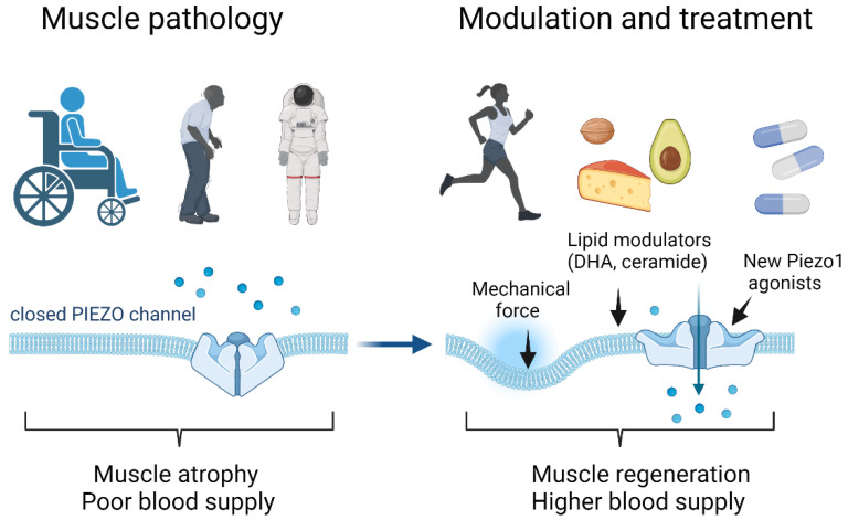
Piezo1 channels are highly mechanically-activated cation channels that can sense and transduce the mechanical stimuli into physiological signals in different tissues including skeletal muscle. In this focused review, we summarize the emerging evidence of Piezo1 channel-mediated effects in the physiology of skeletal muscle, with a particular focus on the role of Piezo1 in controlling myogenic precursor activity and skeletal muscle regeneration and vascularization. The disclosed effects reported by pharmacological activation of Piezo1 channels with the selective agonist Yoda1 indicate a potential impact of Piezo1 channel activity in skeletal muscle regeneration, which is disrupted in various muscular pathological states. All findings reported so far agree with the idea that Piezo1 channels represent a novel, powerful molecular target to develop new therapeutic strategies for preventing or ameliorating skeletal muscle disorders characterized by an impairment of tissue regenerative potential.
Keywords: Piezo1; Yoda1; muscle atrophy; myoblasts; myogenesis; myotubes; sarcopenia; satellite cells.
Conflict of interest statement
The authors declare that they have no conflict of interest.
Figures
Similar articles
-
A Pharmacological Investigation of the TMEM16A Currents in Murine Skeletal Myogenic Precursor Cells.Int J Mol Sci. 2024 Feb 13;25(4):2225. doi: 10.3390/ijms25042225. Int J Mol Sci. 2024. PMID: 38396901 Free PMC article.
-
"Time window" effect of Yoda1-evoked Piezo1 channel activity during mouse skeletal muscle differentiation.Acta Physiol (Oxf). 2021 Dec;233(4):e13702. doi: 10.1111/apha.13702. Epub 2021 Jun 22. Acta Physiol (Oxf). 2021. PMID: 34097801 Free PMC article.
-
Piezo1 channels enhance anabolic signaling activation induced by electrical stimulation of cultured myotubes.FEBS Open Bio. 2025 Jun;15(6):940-948. doi: 10.1002/2211-5463.70008. Epub 2025 Feb 17. FEBS Open Bio. 2025. PMID: 39961145 Free PMC article.
-
The emerging role of Piezo1 channels in skeletal muscle physiology.Biophys Rev. 2023 Sep 29;15(5):1171-1184. doi: 10.1007/s12551-023-01154-6. eCollection 2023 Oct. Biophys Rev. 2023. PMID: 37975010 Free PMC article. Review.
-
Mechanosensitive Cation Channel Piezo1 Is Involved in Renal Fibrosis Induction.Int J Mol Sci. 2024 Jan 31;25(3):1718. doi: 10.3390/ijms25031718. Int J Mol Sci. 2024. PMID: 38338996 Free PMC article. Review.
Cited by
-
A Pharmacological Investigation of the TMEM16A Currents in Murine Skeletal Myogenic Precursor Cells.Int J Mol Sci. 2024 Feb 13;25(4):2225. doi: 10.3390/ijms25042225. Int J Mol Sci. 2024. PMID: 38396901 Free PMC article.
-
Piezo regulates epithelial topology and promotes precision in organ size control.Cell Rep. 2024 Jul 23;43(7):114398. doi: 10.1016/j.celrep.2024.114398. Epub 2024 Jun 26. Cell Rep. 2024. PMID: 38935502 Free PMC article.
-
The molecular athlete: exercise physiology from mechanisms to medals.Physiol Rev. 2023 Jul 1;103(3):1693-1787. doi: 10.1152/physrev.00017.2022. Epub 2023 Jan 5. Physiol Rev. 2023. PMID: 36603158 Free PMC article. Review.
-
Mechanobiology in cellular, molecular, and tissue adaptation.Mechanobiol Med. 2023 Aug 24;1(2):100022. doi: 10.1016/j.mbm.2023.100022. eCollection 2023 Dec. Mechanobiol Med. 2023. PMID: 40395638 Free PMC article.
-
Mechanisms and Countermeasures for Muscle Atrophy in Microgravity.Cells. 2024 Dec 20;13(24):2120. doi: 10.3390/cells13242120. Cells. 2024. PMID: 39768210 Free PMC article. Review.
References
Publication types
MeSH terms
Substances
LinkOut - more resources
Full Text Sources
