. 2022 Jun 14;23(12):6634.
doi: 10.3390/ijms23126634.

Kinase Activity of PAR1b, Which Mediates Nuclear Translocation of the BRCA1 Tumor Suppressor, Is Potentiated by Nucleic Acid-Mediated PAR1b Multimerization

Affiliations

Kinase Activity of PAR1b, Which Mediates Nuclear Translocation of the BRCA1 Tumor Suppressor, Is Potentiated by Nucleic Acid-Mediated PAR1b Multimerization

Hiroko Nishikawa et al. Int J Mol Sci. .

Abstract

PAR1b is a cytoplasmic serine/threonine kinase that controls cell polarity and cell-cell interaction by regulating microtubule stability while mediating cytoplasmic-to-nuclear translocation of BRCA1. PAR1b is also a cellular target of the CagA protein of Helicobacter pylori, which leads to chronic infection causatively associated with the development of gastric cancer. The CagA-PAR1b interaction inactivates the kinase activity of PAR1b and thereby dampens PAR1b-mediated BRCA1 phosphorylation, which reduces the level of nuclear BRCA1 and thereby leads to BRCAness and BRCAness-associated genome instability underlying gastric carcinogenesis. While PAR1b can multimerize within the cells, little is known about the mechanism and functional role of PAR1b multimerization. We found in the present study that PAR1b was multimerized in vitro by binding with nucleic acids (both single- and double-stranded DNA/RNA) via the spacer region in a manner independent of nucleic-acid sequences, which markedly potentiated the kinase activity of PAR1b. Consistent with these in vitro observations, cytoplasmic introduction of double-stranded DNA or expression of single-stranded RNA increased the PAR1b kinase activity in the cells. These findings indicate that the cytoplasmic DNA/RNA contribute to nuclear accumulation of BRCA1 by constitutively activating/potentiating cytoplasmic PAR1b kinase activity, which is subverted in gastric epithelial cells upon delivery of H. pylori CagA oncoprotein.

Keywords: EBER; PAR1b; gastric cancer; kinase activation; multimerization; nucleic acids.


Conflict of interest statement

The authors declare no conflict of interest.

Figures
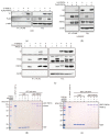
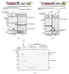
Figure 1
Figure 1
Nucleic acids mediate multimerization of PAR1b. (a) Multimerization of PAR1b in AGS cells. AGS cells were transiently transfected with expression vectors for FLAG-tagged PAR1b and T7-tagged PAR1b. After 24 h, total cell lysates (TCL) were prepared and subjected to immunoprecipitation using the anti-FLAG antibody. Immunoprecipitates (IP) were analyzed by immunoblotting and detected by the indicated antibodies. (b) Multimerization of PAR1b in HEK293T cells. Immunoprecipitation was performed with HEK293T cells transiently transfected with expression vectors for FLAG-tagged PAR1b and HA-tagged PAR1b. (c) Multimerization of PAR1b in AGS cells is mediated by RNA. TCL was treated with RNase A prior to immunoprecipitation. (d) Reconstitution of RNA-mediated multimerization of PAR1b. GST pull-down was performed by mixing GST-PAR1b bound Glutathione Sepharose beads with 100 nM PAR1b in the presence of 1 µg/mL total RNA or mRNA purified from AGS cells. A sample containing only the GST-PAR1b bound beads was analyzed as the input control for GST-PAR1b. (e) DNA can also mediate multimerization of PAR1b. GST pull-down between GST-PAR1b and 100 nM PAR1b was performed in the presence of 1 µg/mL ssDNA or dsDNA prepared from AGS cells. A sample containing only the GST-PAR1b bound beads was analyzed as the input control for GST-PAR1b.
Figure 2
Figure 2
Mapping the multimerization domain of PAR1b. (a) The C-terminus of PAR1b is required for multimerization in cells. Schematic diagram of full-length (FL) FLAG-PAR1b and its N-terminal and C-terminal truncations (top). Amino acid residue numbers refer to the 745 aa isoform of PAR1b (see Methods). AGS cells transiently transfected with expression vectors for the FLAG-PAR1b mutants and T7-PAR1b were subjected to immunoprecipitation (bottom). (b) The spacer region is required for multimerization in cells. Schematic diagram of FLAG-PAR1b-∆spacer (top). AGS cells transiently transfected with the expression vectors for FLAG-PAR1b-∆spacer and T7-PAR1b were subjected to immunoprecipitation (bottom). (c) PARb multimerizes primarily through the spacer region in the presence of nucleic acid. GST-PAR1b-FL bound Glutathione Sepharose beads were mixed with 100 nM PAR1b-FL or PAR1b-∆spacer in the presence of 1 µg/mL total RNA and a GST pull-down assay was performed.
Figure 3
Figure 3
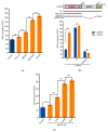
Nucleic acids enhance PAR1b kinase activity. (a) The effect of various nucleic acids on the kinase activity of recombinant PAR1b in an in vitro kinase assay. 200 nM PAR1b-FL was subjected to an in vitro kinase assay in the presence of 40 ng/µL nucleic acids as indicated. Total RNA, ssDNA, and dsDNA were purified from AGS cells. (b) The spacer region is required for the nucleic acid-dependent potentiation of PAR1b kinase activity. 200 nM of the mutant PAR1b proteins indicated (top) were subjected to an in vitro kinase assay in the presence or absence of 40 ng/µL ssDNA from AGS cells (bottom). (c) PAR1b kinase activity is dependent on the length of nucleic acids. 50 nM of PAR1b-FL was subjected to an in vitro kinase assay in the presence of 10 ng/µL ssDNA of different lengths as indicated. Kinase activities were quantified through the amount of ADP produced, which was measured using a fluorescence spectrometer (relative fluorescent units, RFU). Error bars represent mean ± SD, n = 4. * p < 0.05, ** p < 0.01, *** p < 0.001, one-way ANOVA, post-hoc Tukey’s test (a,c) or two-way ANOVA, post-hoc Sidak’s test (b).
Figure 4
Figure 4
PAR1b can multimerize in a nucleic acid-dependent manner in the cell. (a) dsDNA promotes multimerization of PAR1b in cells. AGS cells were transiently transfected with expression vectors for T7-PAR1b and FLAG-PAR1b. After 24 h, cells were transfected with 2 µg/mL 3000 nt ssDNA or 3000 bp dsDNA for 5 h followed by immunoprecipitation. (b) PAR1b can interact with DNA directly through the spacer region in cells. AGS cells were transiently transfected with FLAG-PAR1b expression vectors. After 24 h, cells were transfected with a mixture of pBlueScript II and salmon sperm dsDNA at a ratio of 1:1000 for 2 h before harvest and subsequent DNA-immunoprecipitation. pBlueScript II was detected by PCR using specific primers. (c) Quantitative PCR analysis of DNA immunoprecipitants from (b). Error bars represent mean ± SD, n = 3. *** p < 0.001, one-way ANOVA, post-hoc Tukey’s test.
Figure 5
Figure 5
Nucleic acids can enhance the kinase activity of PAR1b in the cell. (a) Transfection of dsDNA induces phosphorylation of tau at S262 in cells in a time-dependent manner. AGS cells were transiently transfected with an expression vector for FLAG-tau. After 24 h, cells were transfected with 2 µg/mL linearized 3000 bp dsDNA for 2, 4 and 5 h before cell lysis. Cells co-transfected with expression vectors for FLAG-tau and T7-PAR1b are used as positive control (final lane). Phosphorylation of tau was quantified with an image analyzer and normalized to its control at each time point. (b) The effect of various nucleic acid ligands on the phosphorylation of tau. AGS cells were transiently transfected with an expression vector for FLAG-tau followed by transfection with 2 µg/mL of ssDNA from salmon sperm, dsDNA from salmon sperm, total RNA from AGS cells or poly I:C for 2 h. (c) dsDNA induces phosphorylation of tau in AGS cells stably expressing HA-tau. The stable cell line was transfected with 2 µg/mL linearized 3000 bp dsDNA or dsDNA from salmon sperm. (d) Phosphorylation of tau at S262 is dependent on PAR1b. AGS cells stably expressing HA-tau were treated with PAR1b-specific siRNA for 48 h and then transfected with 2 µg/mL dsDNA from salmon sperm for 2 h.
Figure 6
Figure 6
EBERs from Epstein–Barr virus can promote PAR1b multimerization and enhance its kinase activity. (a) Multimerization of PAR1b in AGS cells stably expressing EBERs. AGS cells stably expressing EBERs were transiently transfected with expression vectors for FLAG-tagged PAR1b and HA-tagged PAR1b and immunoprecipitation was performed. (b) Expression of EBERs can potentiate the phosphorylation of tau at S262. HA-tau expression vectors were transiently transfected to three independent clones of AGS cells stably expressing EBERs.

Similar articles

Cited by

References

    1. Marx A., Nugoor C., Panneerselvam S., Mandelkow E. Structure and Function of Polarity-Inducing Kinase Family MARK/Par-1 within the Branch of AMPK/Snf1-Related Kinases. FASEB J. 2010;24:1637–1648. doi: 10.1096/fj.09-148064. - DOI - PubMed
    1. Imai S., Ooki T., Murata-Kamiya N., Komura D., Tahmina K., Wu W., Takahashi-Kanemitsu A., Knight C.T., Kunita A., Suzuki N., et al. Helicobacter pylori CagA Elicits BRCAness to Induce Genome Instability That May Underlie Bacterial Gastric Carcinogenesis. Cell Host Microbe. 2021;29:941–958. doi: 10.1016/j.chom.2021.04.006. - DOI - PubMed
    1. Böhm H., Brinkmann V., Drab M., Henske A., Kurzchalia T.V. Mammalian Homologues of C. elegans PAR-1 Are Asymmetrically Localized in Epithelial Cells and May Influence Their Polarity. Curr. Biol. 1997;7:603–606. doi: 10.1016/S0960-9822(06)00260-0. - DOI - PubMed
    1. Suzuki A., Hirata M., Kamimura K., Maniwa R., Yamanaka T., Mizuno K., Kishikawa M., Hirose H., Amano Y., Izumi N., et al. aPKC Acts Upstream of PAR-1b in Both the Establishment and Maintenance of Mammalian Epithelial Polarity. Curr. Biol. 2004;14:1425–1435. doi: 10.1016/j.cub.2004.08.021. - DOI - PubMed
    1. Drewes G., Trinczek B., Illenberger S., Biernat J., Schmitt-Ulms G., Meyer H.E., Mandelkow E.-M., Mandelkow E. Microtubule-Associated Protein/Microtubule Affinity-Regulating Kinase (P110mark): A Novel Protein Kinase That Regulates Tau-Microtubule Interactions and Dynamic Instability by Phosphorylation at the Alzheimer-Specific Site Serine 262. J. Biol. Chem. 1995;270:7679–7688. doi: 10.1074/jbc.270.13.7679. - DOI - PubMed