. 2022 Jun 16;23(12):6705.
doi: 10.3390/ijms23126705.

NMDA and P2X7 Receptors Require Pannexin 1 Activation to Initiate and Maintain Nociceptive Signaling in the Spinal Cord of Neuropathic Rats

Affiliations

NMDA and P2X7 Receptors Require Pannexin 1 Activation to Initiate and Maintain Nociceptive Signaling in the Spinal Cord of Neuropathic Rats

David Bravo et al. Int J Mol Sci. .

Abstract

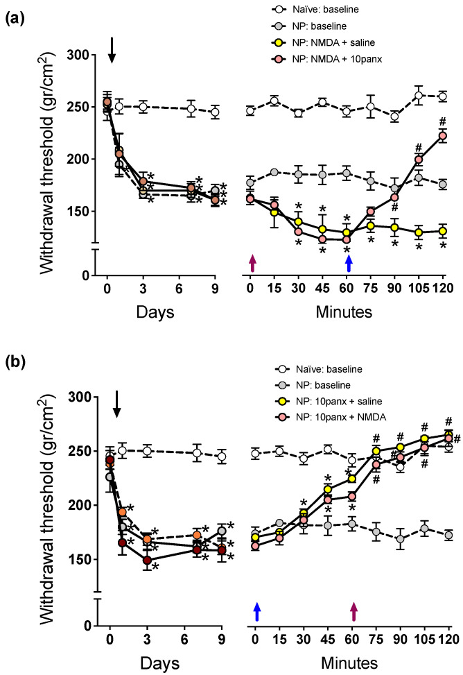
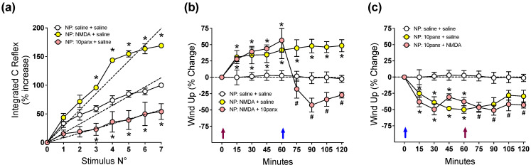
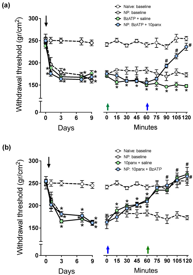
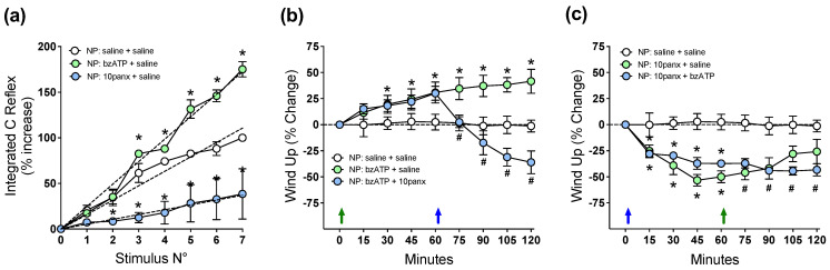
Pannexin 1 (Panx1) is involved in the spinal central sensitization process in rats with neuropathic pain, but its interaction with well-known, pain-related, ligand-dependent receptors, such as NMDA receptors (NMDAR) and P2X7 purinoceptors (P2X7R), remains largely unexplored. Here, we studied whether NMDAR- and P2X7R-dependent nociceptive signaling in neuropathic rats require the activation of Panx1 channels to generate spinal central sensitization, as assessed by behavioral (mechanical hyperalgesia) and electrophysiological (C-reflex wind-up potentiation) indexes. Administration of either a selective NMDAR agonist i.t. (NMDA, 2 mM) or a P2X7R agonist (BzATP, 150 μM) significantly increased both the mechanical hyperalgesia and the C-reflex wind-up potentiation, effects that were rapidly reversed (minutes) by i.t. administration of a selective pannexin 1 antagonist (10panx peptide, 300 μM), with the scores even reaching values of rats without neuropathy. Accordingly, 300 μM 10panx completely prevented the effects of NMDA and BzATP administered 1 h later, on mechanical hyperalgesia and C-reflex wind-up potentiation. Confocal immunofluorescence imaging revealed coexpression of Panx1 with NeuN protein in intrinsic dorsal horn neurons of neuropathic rats. The results indicate that both NMDAR- and P2X7R-mediated increases in mechanical hyperalgesia and C-reflex wind-up potentiation require neuronal Panx1 channel activation to initiate and maintain nociceptive signaling in neuropathic rats.

Keywords: NMDA receptor; P2X7 receptor; neuropathic pain; pannexin 1; wind-up.


Conflict of interest statement

The authors declare no conflict of interest.

Figures

Figure 1
Figure 1
Effect of intrathecal administration of NMDA followed by 10panx, or 10panx followed by NMDA, on the mechanical nociceptive threshold (in g/cm2) of neuropathic (NP) rats. Naïve (baseline) and NP (baseline) rats without receiving any drug served as controls. Values are means ± SEM, n = 6 rats in each group. (a) Left panel: Time course of development of NP after sural nerve cutting, performed on day zero (downward arrow), during a 9-day period of follow-up. Two-way repeated measures ANOVA revealed a “time” effect (F2.173,43.46 = 53.40). Intragroup analysis: * p at least <0.05 in NP rats with respect to the threshold prior to sural nerve cutting, Bonferroni multiple comparisons test. Right panel: Time course of changes in mechanical nociceptive threshold on day 10, after a 10 µL injection of saline i.t. or 2 mM NMDA i.t. at time zero min (violet upward arrow), followed by saline i.t. or 300 µM 10panx i.t. at time 60 min (blue upward arrow). Two-way repeated measures ANOVA revealed a “time” effect (F5.282,105.6 = 15.03). Intragroup analysis: * p at least <0.05 with respect to time zero min, # p at least <0.05 with respect to time 60 min, Bonferroni multiple comparisons test). Note the hyperalgesic effect of NMDA, and the counteracting effect of 10panx. (b) Left panel: Time course of development of NP after sural nerve cutting, performed on day zero (downward arrow), during a 9-day period of follow-up. Two-way repeated measures ANOVA revealed a “time” effect (F3.036,60.71 = 54.54). Intragroup analysis: * p at least <0.05 in NP rats with respect to the threshold prior to sural nerve cutting, Bonferroni multiple comparisons test. Right panel: Time course of changes in the mechanical nociceptive threshold of NP rats on day 10, upon an inverse scheme of drug administration: a 10 µL injection of saline i.t. or 300 µM 10panx i.t. was administered at time zero min (blue arrow), and saline i.t. or 2 mM NMDA i.t. one hour later, at time 60 min (violet arrow). Two-way repeated measures ANOVA revealed a “time” effect (F5.455,109.1 = 48.38). Intragroup analysis: * p at least <0.05 with respect to time zero min, # p at least <0.05 with respect to time 60 min, Bonferroni multiple comparisons test. Note the antihyperalgesic effect of 10panx, and its preventing effect on the ability of NMDA to induce hyperalgesia.
Figure 2
Figure 2
Effect of intrathecal administration of NMDA followed by 10panx, or 10panx followed by NMDA, on C-reflex wind-up activity of neuropathic (NP) rats. NP rats receiving saline served as controls. Values are means ± SEM, n = 6 rats in each group. (a) Figure depicting data processing for determining spinal cord wind-up scores in neuropathic animals. C-reflex responses were elicited by repetitive electric stimulation (1 Hz) of the second and third toes to develop wind-up potentiation, a frequency-dependent increase in the excitability of spinal cord neurons. The C-reflex responses were integrated and plotted against the stimulus number, and the curves were normalized so that the reflex gain at the seventh stimulus represented a 100% increase. The slope of the least-squares regression line represents a control wind-up score, prior to any administration of drugs or saline. Thereafter, a 10 µL i.t. injection of either saline solution (white circles), 2 mM NMDA (yellow circles) or 300 µM 10panx (pink circles) was performed, and a new series of seven repetitive electric stimulations (1 Hz) was applied 30 min after, and the wind-up scores calculated from the slopes of the respective least-squares regression lines. The procedure was repeated using 6 animals per group, and the average data is presented as Figure a. Values are means ± SEM of data averaged from 6 rats per group, and the slopes of regression curves (dashed lines) represent wind-up scores obtained 30 min after each drug treatment: 16.15 ± 0.70 (saline), 28.48 ± 0.99 * (NMDA), and 8.14 ± 0.95* (10panx), the difference between slopes being statistically significant (one-way ANOVA, F2,15 = 132.7; * p at least <0.0001 with respect to saline group, Bonferroni multiple comparisons test). (b) Time course of wind-up activity (expressed as % change) in neuropathic rats immediately before and 15, 30, 45, 60, 75, 90, 105, and 120 min after an i.t. injection of NMDA or saline at time 0 (violet arrow), followed by an i.t. injection of 10panx or saline at time 60 min (blue arrow). Two-way repeated measures ANOVA revealed a “time” effect (F1.732,29.44 = 4.504). Intragroup analysis: * p at least <0.05 with respect to time zero min, # p at least <0.05 with respect to time 60 min, Bonferroni multiple comparisons test). Note the increased wind-up activity after MDMA, and the counteracting effect of 10panx. (c) Time course of wind-up activity (% change) in neuropathic rats after the inverse scheme of drug administration, i.e., an i.t. injection of 10panx or saline at time 0 (blue arrow), followed by an i.t. injection of NMDA or saline at time 60 min (violet arrow). Two-way repeated measures ANOVA revealed a “time” effect (F4.717,70.75 = 8.484). Intragroup analysis: * p at least <0.05 with respect to time zero min, # p at least <0.05 with respect to time 60 min, Bonferroni multiple comparisons test). Note the decreased wind-up activity after 10panx, and its preventing effect on the ability of NMDA to increase wind-up.
Figure 3
Figure 3
Effect of intrathecal administration of BzATP followed by 10panx, or 10panx followed by BzATP, on the mechanical nociceptive threshold (in g/cm2) of neuropathic (NP) rats. Naïve (baseline) and NP (baseline) rats without receiving any drug served as controls. Values are means ± SEM, n = 6 rats in each group. (a) Left panel: Time course of development of NP after sural nerve cutting, performed on day zero (downward arrow), during a 9-day period of follow-up. Two-way repeated measures ANOVA revealed a “time” effect (F2.992,59.84 = 65.12). Intragroup analysis: * p at least <0.05 in NP rats with respect to the threshold prior to sural nerve cutting, Bonferroni multiple comparisons test. Right panel: Time course of changes in mechanical nociceptive threshold after a 10 µL injection of saline i.t. or 150 µM BzATP i.t. on day 10 at time zero min (green upward arrow), followed by saline i.t. or 300 µM 10panx i.t. at time 60 min (blue upward arrow). Two-way repeated measures ANOVA revealed a “time” effect (F5.628,112.6 = 8.096). Intragroup analysis: * p at least <0.05 with respect to time zero min, # p at least <0.05 with respect to time 60 min, Bonferroni multiple comparisons test). Note the hyperalgesic effect of NMDA, and the counteracting effect of 10panx. (b) Left panel: Time course of development of NP after sural nerve cutting, performed on day zero (downward arrow), during a 9-day period of follow-up. Two-way repeated measures ANOVA revealed a “time” effect (F2.112, 42.24 = 50.46). Intragroup analysis: * p at least <0.05 in NP rats with respect to the threshold prior to sural nerve cutting, Bonferroni multiple comparisons test. Right panel: Time course of changes in the mechanical nociceptive threshold of NP rats on an inverse scheme of drug administration: a 10 µL injection of saline i.t. or 300 µM 10panx i.t. was administered at time zero min (blue arrow), and saline i.t. or 150 µM BzATP i.t. one hour later, at time 60 min (green arrow). Two-way repeated measures ANOVA revealed a “time” effect (F5.437,108.7 = 53.94). Intragroup analysis: * p at least <0.05 with respect to time zero min, # p at least <0.05 with respect to time 60 min, Bonferroni multiple comparisons test). Note the antihyperalgesic effect of 10panx, and its preventing effect on the ability of BzATP to induce hyperalgesia.
Figure 4
Figure 4
Effect of intrathecal administration of BzATP followed by 10panx, or 10panx followed by BzATP, on C-reflex wind-up activity of neuropathic (NP) rats. NP rats receiving saline served as controls. Values are means ± SEM, n = 6 rats in each group. (a) Figure depicting data processing for determining spinal cord wind-up scores in neuropathic animals, as in Figure 2a. The slope of the least-squares regression line represents a control wind-up score, prior to any administration of drugs or saline. Thereafter, a 10 µL i.t. injection of either saline solution (white circles), 150 µM BzATP (light green circles) or 300 µM 10panx (light blue circles) was performed, and a new series of seven repetitive electric stimulations (1 Hz) was applied 30 min after, and the wind-up scores calculated from the slopes of the respective least-squares regression lines. The procedure was repeated using 6 animals per group, and the average data is presented as Figure 4a. Values are means ± SEM of data averaged from 6 rats per group, and the slopes of regression curves (dashed lines) represent wind-up scores for each drug treatment: 15.80 ± 0.58 (saline), 24.62 ± 0.58 * (BzATP), and 5.28 ± 0.63 * (10panx), the difference between slopes being statistically significant (one-way ANOVA, F2,15 = 378.5; * p at least <0.0001 with respect to saline group, Bonferroni multiple comparisons test). (b) Time course of wind-up activity (expressed as % change) in neuropathic rats immediately before and 15, 30, 45, 60, 75, 90, 105 and 120 min after an i.t. injection of BzATP or saline at time 0 (violet arrow), followed by an i.t. injection of 10panx or saline at time 60 min (blue arrow). Two-way repeated measures ANOVA revealed a “time” effect (F1.386,20.78 = 3.701). Intragroup analysis: * p at least <0.05 with respect to time zero min, # p at least <0.05 with respect to time 60 min, Bonferroni multiple comparisons test). Note the increased wind-up activity after BzATP, and the counteracting effect of 10panx. (c) Time course of wind-up activity (% change) in neuropathic rats after the inverse scheme of drug administration, i.e., an i.t. injection of 10panx or saline at time 0 (blue arrow), followed by an i.t. injection of BzATP or saline at time 60 min (violet arrow). Two-way repeated measures ANOVA revealed a “time” effect (F3.877,58.15 = 8.480). Intragroup analysis: * p at least <0.05 with respect to time zero min, # p at least <0.05 with respect to time 60 min, Bonferroni multiple comparisons test. Note the decreased wind-up activity after 10panx, and its preventing effect on the ability of BzATP to increase wind-up.
Figure 5
Figure 5
Confocal images from a 200µm-thick lumbar spinal cord transverse slice of a neuropathic rat, showing some selected neurons from Rexed laminae I and II (see white arrows) labeled with NeuN immunofluorescence (left panel), Panx1 immunofluorescence (middle panel), and double Neun/Panx1 immunofluorescence (right panel). Panx1 channel expression was often visualized surrounding neuronal nuclei colabeled with the NeuN protein, thus indicating the presence of Panx1 in intrinsic dorsal horn neurons. Blue labeling in the right panel corresponds to DAPI fluorescent nuclear stain. Scale bars: 50 µm in all three panels.

Similar articles

Cited by

References

    1. Latremoliere A., Woolf C.J. Central Sensitization: A Generator of Pain Hypersensitivity by Central Neural Plasticity. J. Pain. 2009;10:895–926. doi: 10.1016/j.jpain.2009.06.012. - DOI - PMC - PubMed
    1. Kuner R. Central mechanisms of pathological pain. Nat. Med. 2010;16:1258–1266. doi: 10.1038/nm.2231. - DOI - PubMed
    1. Bannister K., Sachau J., Baron R., Dickenson A.H. Neuropathic Pain: Mechanism-Based Therapeutics. Annu. Rev. Pharmacol. Toxicol. 2020;60:257–274. doi: 10.1146/annurev-pharmtox-010818-021524. - DOI - PubMed
    1. Inoue K., Tsuda M. Purinergic systems, neuropathic pain and the role of microglia. Exp. Neurol. 2011;234:293–301. doi: 10.1016/j.expneurol.2011.09.016. - DOI - PubMed
    1. Ji R.-R., Berta T., Nedergaard M. Glia and pain: Is chronic pain a gliopathy? Pain. 2013;154:S10–S28. doi: 10.1016/j.pain.2013.06.022. - DOI - PMC - PubMed

LinkOut - more resources