. 2022 Jun 17;23(12):6769.
doi: 10.3390/ijms23126769.

Identification of 8-Hydroxyquinoline Derivatives That Decrease Cystathionine Beta Synthase (CBS) Activity

Affiliations

Identification of 8-Hydroxyquinoline Derivatives That Decrease Cystathionine Beta Synthase (CBS) Activity

Pierre Conan et al. Int J Mol Sci. .

Abstract

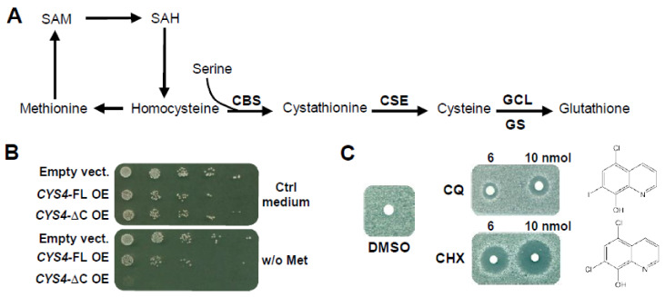
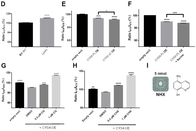
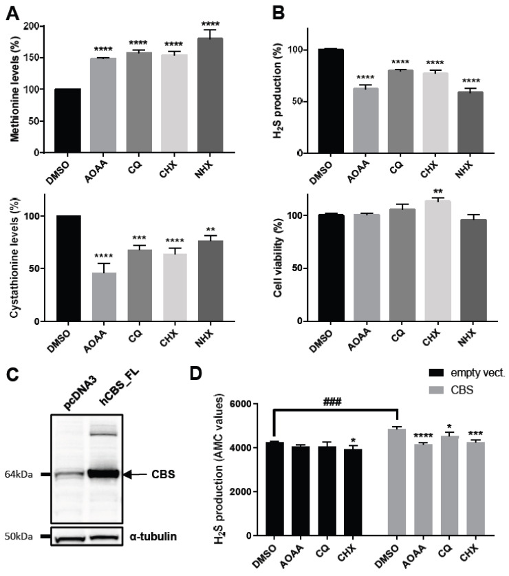
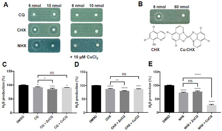
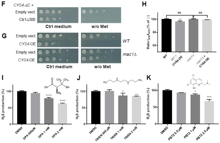
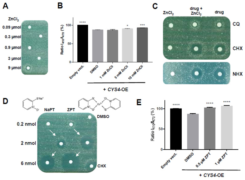
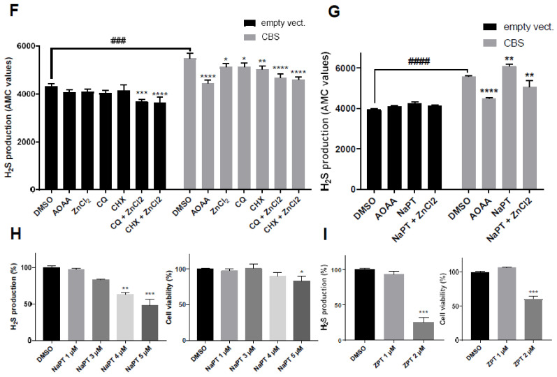
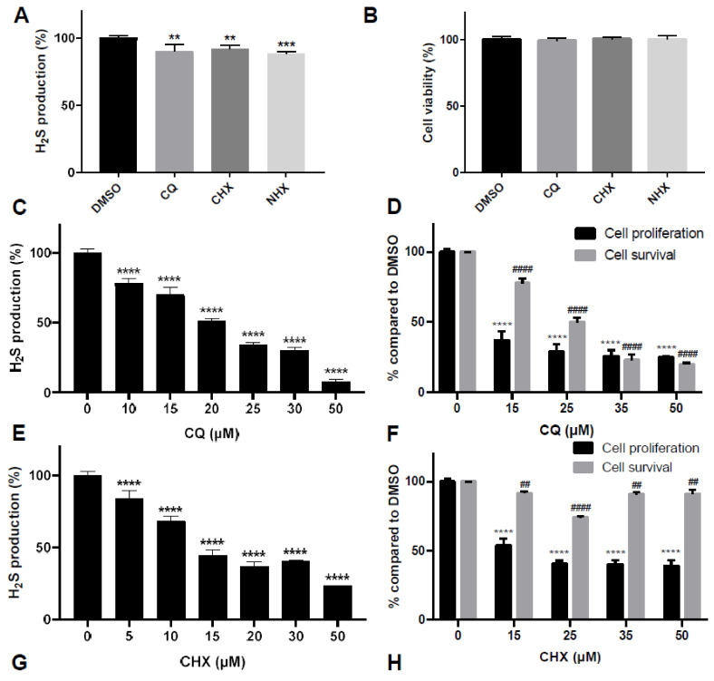
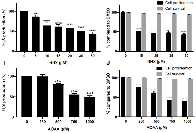
CBS encodes a pyridoxal 5'-phosphate-dependent enzyme that catalyses the condensation of homocysteine and serine to form cystathionine. Due to its implication in some cancers and in the cognitive pathophysiology of Down syndrome, the identification of pharmacological inhibitors of this enzyme is urgently required. However, thus far, attempts to identify such molecules have only led to the identification of compounds with low potency and limited selectivity. We consequently developed an original, yeast-based screening method that identified three FDA-approved drugs of the 8-hydroxyquinoline family: clioquinol, chloroxine and nitroxoline. These molecules reduce CBS enzymatic activity in different cellular models, proving that the molecular mechanisms involved in yeast phenotypic rescue are conserved in mammalian cells. A combination of genetic and chemical biology approaches also revealed the importance of copper and zinc intracellular levels in the regulation of CBS enzymatic activity-copper promoting CBS activity and zinc inhibiting its activity. Taken together, these results indicate that our effective screening approach identified three new potent CBS inhibitors and provides new findings for the regulation of CBS activity, which is crucial to develop new therapies for CBS-related human disorders.

Keywords: CBS; Cys4; Gex1/Gex2; copper; cytosolic pH; drug screening; zinc.


Conflict of interest statement

The authors have no relevant financial or non-financial interests to disclose.

Figures

Figure 1
Figure 1
Identification of molecules decreasing the effects of CYS4 overexpression (CYS4-OE) in yeast. (A) Simplified representation of the trans-sulphuration pathway. In yeast, CYS4 encodes the cystathionine beta synthase protein (CBS or Cys4p in yeast), which converts homocysteine and serine into cystathionine. The other enzymes of this pathway are cystathionine gamma-lyase (CSE), (γ-glutamylcysteine synthetase (GCL) and glutathione synthetase (GS). CYS4 is located at a metabolic hub, its deletion leading to decreased synthesis of cysteine and glutathione in favour of S-adenosylmethionine (SAM), S-adenosylhomocysteine (SAH) and methionine synthesis, whereas CYS4-OE favours cysteine and glutathione synthesis at the expense of methionine. (B) Methionine auxotrophy of CYS4-OE cells. Methionine auxotrophy, revealed by the absence of growth on a methionine-free medium, was assessed by spotting serial dilutions of wildtype yeast cells transformed with two 2µ plasmids either empty or containing full-length (CYS4-FL) or a truncated form of CYS4 (CYS4-ΔC), the expression of which is driven by the strong GPD promoter. No serine was added here in order to see the increased methionine auxotrophy obtained with CYS4-ΔC compared to CYS4-FL. (C) Clioquinol (CQ) and chloroxine (CHX) rescue the growth of CYS4-OE yeast cells on a methionine-free medium. The indicated amount of drug was added on filters. The positive action of the drugs on cell growth is indicated by the presence of a clear halo surrounding the filters where the molecules were deposited. Due to the gradient effect of the drug deposited on filters, note that these two molecules are toxic at very high concentrations (dark halos around the filters) but are active at lower concentrations (white halos corresponding to the rescue of cell growth). (DF) Effect of CYS4 deletion or OE on cytosolic pH assessed using a pHluorin plasmid. Quantitative measurements of the I410/I470 ratio of fluorescence showed an increased ratio for cys4Δ cells (D), indicative of an increased cytosolic pH and a decreased ratio of fluorescence for wildtype cells overexpressing either form of Cys4p (full-length or C-terminal domain deleted) (E), indicative of reduced cytosolic pH. Addition of 1.5 mM of serine in the medium of CYS4-OE cells also further decreased cytosolic pH (F), showing the existence of a direct link between Cys4p enzymatic activity and cytosolic acidification. (G,H) Dose-dependent rescue of cytosolic acidification of CYS4-OE cells by CQ (G) and CHX (H). Note that at 1 µM, CQ and CHX even increased cytosolic pH above the level of control cells, which is probably the result of their pro-oxidant effect in certain growth conditions, which may partly deplete intracellular glutathione and/or inhibit endogenous Cys4p. (I) Nitroxoline (NHX) rescues the growth of CYS4-OE yeast cells on a methionine-free medium. Note that NHX was toxic at 10 nmol. (D) Student’s t-test. (E,F) One-way ANOVA with Tukey’s post-hoc test. (G,H): Comparison of each condition with DMSO, one-way ANOVA with Dunnett’s post-hoc test. *, p < 0.05; **, p < 0.01; ***, p < 0.001; ****, p < 0.0001.
Figure 1
Figure 1
Identification of molecules decreasing the effects of CYS4 overexpression (CYS4-OE) in yeast. (A) Simplified representation of the trans-sulphuration pathway. In yeast, CYS4 encodes the cystathionine beta synthase protein (CBS or Cys4p in yeast), which converts homocysteine and serine into cystathionine. The other enzymes of this pathway are cystathionine gamma-lyase (CSE), (γ-glutamylcysteine synthetase (GCL) and glutathione synthetase (GS). CYS4 is located at a metabolic hub, its deletion leading to decreased synthesis of cysteine and glutathione in favour of S-adenosylmethionine (SAM), S-adenosylhomocysteine (SAH) and methionine synthesis, whereas CYS4-OE favours cysteine and glutathione synthesis at the expense of methionine. (B) Methionine auxotrophy of CYS4-OE cells. Methionine auxotrophy, revealed by the absence of growth on a methionine-free medium, was assessed by spotting serial dilutions of wildtype yeast cells transformed with two 2µ plasmids either empty or containing full-length (CYS4-FL) or a truncated form of CYS4 (CYS4-ΔC), the expression of which is driven by the strong GPD promoter. No serine was added here in order to see the increased methionine auxotrophy obtained with CYS4-ΔC compared to CYS4-FL. (C) Clioquinol (CQ) and chloroxine (CHX) rescue the growth of CYS4-OE yeast cells on a methionine-free medium. The indicated amount of drug was added on filters. The positive action of the drugs on cell growth is indicated by the presence of a clear halo surrounding the filters where the molecules were deposited. Due to the gradient effect of the drug deposited on filters, note that these two molecules are toxic at very high concentrations (dark halos around the filters) but are active at lower concentrations (white halos corresponding to the rescue of cell growth). (DF) Effect of CYS4 deletion or OE on cytosolic pH assessed using a pHluorin plasmid. Quantitative measurements of the I410/I470 ratio of fluorescence showed an increased ratio for cys4Δ cells (D), indicative of an increased cytosolic pH and a decreased ratio of fluorescence for wildtype cells overexpressing either form of Cys4p (full-length or C-terminal domain deleted) (E), indicative of reduced cytosolic pH. Addition of 1.5 mM of serine in the medium of CYS4-OE cells also further decreased cytosolic pH (F), showing the existence of a direct link between Cys4p enzymatic activity and cytosolic acidification. (G,H) Dose-dependent rescue of cytosolic acidification of CYS4-OE cells by CQ (G) and CHX (H). Note that at 1 µM, CQ and CHX even increased cytosolic pH above the level of control cells, which is probably the result of their pro-oxidant effect in certain growth conditions, which may partly deplete intracellular glutathione and/or inhibit endogenous Cys4p. (I) Nitroxoline (NHX) rescues the growth of CYS4-OE yeast cells on a methionine-free medium. Note that NHX was toxic at 10 nmol. (D) Student’s t-test. (E,F) One-way ANOVA with Tukey’s post-hoc test. (G,H): Comparison of each condition with DMSO, one-way ANOVA with Dunnett’s post-hoc test. *, p < 0.05; **, p < 0.01; ***, p < 0.001; ****, p < 0.0001.
Figure 2
Figure 2
Molecules reducing the phenotypes induced by CYS4-OE-induced a decrease in human CBS activity. (A) Effects of CQ, CHX and NHX on the intracellular levels of methionine (upper panel) and cystathionine (lower panel). A 24 h treatment of HepG2 cells with 500 µM of AOAA, used as a positive control, and 15 µM of CQ, CHX or NHX resulted in decreased intracellular levels of methionine and increased levels of cystathionine compared to cells treated with the vehicle (DMSO). (B) Effects of CQ, CHX and NHX on H2S production in HepG2 cells. H2S production and cell viability were assessed by AzMC probe and WST-8 assay, respectively. A 24 h treatment of HepG2 cells with 500 µM of AOAA and 20 µM of CQ, CHX or NHX resulted in decreased H2S production (upper panel) without decreasing cell viability (lower panel). Note that CHX tended to increase cell proliferation at 20 µM (see also Figure S3). (C) Western blot showing the level of expression of CBS in cellular lysates of HepG2 cells transfected with the expression plasmid pcDNA3-hCBS compared to cells transfected with the pcDNA3 empty vector. (D) Measure of H2S production in cell lysates obtained from pcDNA3 (empty vector) or CBS-transfected cells. Each molecule was incubated at 50 µM with the indicated cell lysate for 2 h, and the level of H2S production was assessed by AzMC probe. Note that in CBS-transfected cells, AOAA decreases the level of H2S production to that obtained in control cells. Student’s t-test: ###, p < 0.001. (A,B,D) Comparison of each condition with DMSO, one-way ANOVA with Dunnett’s post-hoc test: *, p < 0.05; **, p < 0.01; ***, p < 0.001, ****, p < 0.0001.
Figure 3
Figure 3
Decreasing intracellular copper levels reduces cystathionine beta synthase enzymatic activity. (A) Effect of copper enrichment in the medium. CYS4-OE cells were grown on a methionine-free medium containing 1.5 mM of serine (left panel). Addition of 10 µM of CuCl2 in the medium (right panel) completely abolished the effect of CQ, CHX or NHX on growth restoration of CYS4-OE cells. Note that the cellular toxicity of the drugs at high doses (indicated by dark halos around the filters) was also completely abolished by CuCl2. (B) Test of CHX complexed to copper. A CHX-copper complex (filter on the right side of the plate) was unable to restore cell growth of CYS4-OE cells on a free-methionine medium, in contrast to CHX alone (filter on the left). (CE) Effect of a 24 h incubation of HepG2 cells with a combination of the drug with zinc salts or copper salts on H2S production. Addition of 2.5 µM of CuCl2 did not have any significant effect on the action of 10 µM of CQ (C) or CHX (D) but significantly increased the toxicity of NHX ((E) and Figure S4C–E). The addition of 10 µM of ZnCl2 slightly increased CQ (C) or CHX (D) activity but did not have any effect on NHX (E). Higher concentrations of ZnCl2 or CuCl2 combined with CQ, CHX or NHX decreased cell viability (data not shown). (F) Effect of intracellular copper enrichment on the methionine auxotrophy phenotype of CYS4-OE cells. Expression of Ctr1Δ300 (leading to increased intracellular copper levels) exacerbated methionine auxotrophy. This assay was performed in a yeast strain transfected with only one vector expressing CYS4-ΔC instead of two in order to have an intermediate stringency of methionine auxotrophy due to CYS4-OE. Strains expressing Ctr1Δ300 or control empty plasmids were spotted in serial dilutions on control medium containing methionine (left panel) and on a methionine-free medium (right panel) to assess their growth. (G,H) Effect of intracellular copper depletion on cellular phenotypes of CYS4-OE cells. (G) MAC1 encodes a transcription factor activating the expression of copper transporters in yeast, and its deletion has been shown to induce intracellular copper depletion. In the absence of MAC1, CYS4-OE did not induce methionine auxotrophy. (H) Similarly, CYS4-OE was not able to induce cytosolic acidification in a Mac1Δ strain. (IK) Effect of copper chelators on H2S production in HepG2 cells. A 24 h incubation with three copper chelators, D-penicillamine (I), trientine (J) and PBT2 (K), resulted in decreased H2S production in HepG2 cells. Cell viability was assessed by WST-8 assay (Figure S4F–H). (CE,H) One-way ANOVA with Tukey’s post-hoc test. (IK) Comparison of each condition with DMSO, one-way ANOVA with Dunnett’s post-hoc test. ns: not statistically significant; *, p < 0.05; **, p < 0.01; ***, p < 0.001, ****, p < 0.0001.
Figure 3
Figure 3
Decreasing intracellular copper levels reduces cystathionine beta synthase enzymatic activity. (A) Effect of copper enrichment in the medium. CYS4-OE cells were grown on a methionine-free medium containing 1.5 mM of serine (left panel). Addition of 10 µM of CuCl2 in the medium (right panel) completely abolished the effect of CQ, CHX or NHX on growth restoration of CYS4-OE cells. Note that the cellular toxicity of the drugs at high doses (indicated by dark halos around the filters) was also completely abolished by CuCl2. (B) Test of CHX complexed to copper. A CHX-copper complex (filter on the right side of the plate) was unable to restore cell growth of CYS4-OE cells on a free-methionine medium, in contrast to CHX alone (filter on the left). (CE) Effect of a 24 h incubation of HepG2 cells with a combination of the drug with zinc salts or copper salts on H2S production. Addition of 2.5 µM of CuCl2 did not have any significant effect on the action of 10 µM of CQ (C) or CHX (D) but significantly increased the toxicity of NHX ((E) and Figure S4C–E). The addition of 10 µM of ZnCl2 slightly increased CQ (C) or CHX (D) activity but did not have any effect on NHX (E). Higher concentrations of ZnCl2 or CuCl2 combined with CQ, CHX or NHX decreased cell viability (data not shown). (F) Effect of intracellular copper enrichment on the methionine auxotrophy phenotype of CYS4-OE cells. Expression of Ctr1Δ300 (leading to increased intracellular copper levels) exacerbated methionine auxotrophy. This assay was performed in a yeast strain transfected with only one vector expressing CYS4-ΔC instead of two in order to have an intermediate stringency of methionine auxotrophy due to CYS4-OE. Strains expressing Ctr1Δ300 or control empty plasmids were spotted in serial dilutions on control medium containing methionine (left panel) and on a methionine-free medium (right panel) to assess their growth. (G,H) Effect of intracellular copper depletion on cellular phenotypes of CYS4-OE cells. (G) MAC1 encodes a transcription factor activating the expression of copper transporters in yeast, and its deletion has been shown to induce intracellular copper depletion. In the absence of MAC1, CYS4-OE did not induce methionine auxotrophy. (H) Similarly, CYS4-OE was not able to induce cytosolic acidification in a Mac1Δ strain. (IK) Effect of copper chelators on H2S production in HepG2 cells. A 24 h incubation with three copper chelators, D-penicillamine (I), trientine (J) and PBT2 (K), resulted in decreased H2S production in HepG2 cells. Cell viability was assessed by WST-8 assay (Figure S4F–H). (CE,H) One-way ANOVA with Tukey’s post-hoc test. (IK) Comparison of each condition with DMSO, one-way ANOVA with Dunnett’s post-hoc test. ns: not statistically significant; *, p < 0.05; **, p < 0.01; ***, p < 0.001, ****, p < 0.0001.
Figure 4
Figure 4
Effect of zinc enrichment on cystathionine beta synthase activity. (AE) Effect of zinc enrichment of the medium on the phenotypes of CYS4-OE cells. (A) CYS4-OE cells were grown on a methionine-free medium, and increasing amounts of ZnCl2 were loaded on filters. At the highest dose tested (9 µmol), ZnCl2 on its own was sufficient to restore cell growth of CYS4-OE cells on a methionine-free medium. (B) Addition of increasing amounts of ZnCl2 increased the intracellular pH of CYS4-OE cells in a dose-dependent manner. (C) CYS4-OE cells were grown on a methionine-free medium, and 0.3 µmol of ZnCl2 was added either alone (left filter) or in combination with the tested drug (middle filter). Compared to the drug alone (right filter; 6 nmol of CQ, CHX or NHX), the addition of ZnCl2 slightly increased the activity of CQ and CHX, as shown by their increased toxicity and the decreased concentrations of the molecule necessary to restore cell growth on a methionine-free medium (indicated by the larger distance of the halo from the filter). However, ZnCl2 did not appear to have any obvious effect on NHX action, which is consistent with published findings that NHX is not a zinc ionophore. (D) Pyrithione, a zinc ionophore, was able to rescue cell growth of CYS4-OE cells on a methionine-free medium. Note that zinc pyrithione (ZPT) was more active and more toxic compared to the same amounts of sodium pyrithione (NaPT). (E) Similarly, zinc pyrithione also increased the cytosolic pH of CYS4-OE cells. (F) Effect of the addition of ZnCl2 to CHX and CQ on H2S production in cell lysates. The effect of 50 µM of CQ or CHX on H2S production was visible after a 2 h incubation with cell lysates obtained from pcDNA3- or CBS-transfected HepG2 cells. The efficiency of both CQ and CHX was more pronounced in CBS-transfected cells, which produced higher levels of H2S compared to control-transfected cells. The action of CQ and CHX was also enhanced by 50 µM of ZnCl2 in cell lysates. AOAA (50 µM) was used as a positive control. (G) Effect of the addition of ZnCl2 on NaPT in cell lysates. Whereas the addition of 50 µM of NaPT on its own increased the production of H2S, the addition of 50 µM of ZnCl2 to 50 µM of NaPT significantly decreased the production of H2S after a 2 h incubation with cell lysates obtained from CBS-transfected HepG2 cells. (H,I) Dose-dependent effect of pyrithione complexed to sodium (NaPT) or to zinc (ZPT) on H2S production after a 24 h incubation of HepG2 cells. Cell viability (right panel) was assessed by WST-8 assay. (B,EI) Comparison of each condition with DMSO, one-way ANOVA with Dunnett’s post-hoc test: *, p < 0.05; **, p < 0.01; ***, p < 0.001, ****, p < 0.0001. (F,G) Student’s t-test: ###, p < 0.001, ####, p < 0.0001.
Figure 4
Figure 4
Effect of zinc enrichment on cystathionine beta synthase activity. (AE) Effect of zinc enrichment of the medium on the phenotypes of CYS4-OE cells. (A) CYS4-OE cells were grown on a methionine-free medium, and increasing amounts of ZnCl2 were loaded on filters. At the highest dose tested (9 µmol), ZnCl2 on its own was sufficient to restore cell growth of CYS4-OE cells on a methionine-free medium. (B) Addition of increasing amounts of ZnCl2 increased the intracellular pH of CYS4-OE cells in a dose-dependent manner. (C) CYS4-OE cells were grown on a methionine-free medium, and 0.3 µmol of ZnCl2 was added either alone (left filter) or in combination with the tested drug (middle filter). Compared to the drug alone (right filter; 6 nmol of CQ, CHX or NHX), the addition of ZnCl2 slightly increased the activity of CQ and CHX, as shown by their increased toxicity and the decreased concentrations of the molecule necessary to restore cell growth on a methionine-free medium (indicated by the larger distance of the halo from the filter). However, ZnCl2 did not appear to have any obvious effect on NHX action, which is consistent with published findings that NHX is not a zinc ionophore. (D) Pyrithione, a zinc ionophore, was able to rescue cell growth of CYS4-OE cells on a methionine-free medium. Note that zinc pyrithione (ZPT) was more active and more toxic compared to the same amounts of sodium pyrithione (NaPT). (E) Similarly, zinc pyrithione also increased the cytosolic pH of CYS4-OE cells. (F) Effect of the addition of ZnCl2 to CHX and CQ on H2S production in cell lysates. The effect of 50 µM of CQ or CHX on H2S production was visible after a 2 h incubation with cell lysates obtained from pcDNA3- or CBS-transfected HepG2 cells. The efficiency of both CQ and CHX was more pronounced in CBS-transfected cells, which produced higher levels of H2S compared to control-transfected cells. The action of CQ and CHX was also enhanced by 50 µM of ZnCl2 in cell lysates. AOAA (50 µM) was used as a positive control. (G) Effect of the addition of ZnCl2 on NaPT in cell lysates. Whereas the addition of 50 µM of NaPT on its own increased the production of H2S, the addition of 50 µM of ZnCl2 to 50 µM of NaPT significantly decreased the production of H2S after a 2 h incubation with cell lysates obtained from CBS-transfected HepG2 cells. (H,I) Dose-dependent effect of pyrithione complexed to sodium (NaPT) or to zinc (ZPT) on H2S production after a 24 h incubation of HepG2 cells. Cell viability (right panel) was assessed by WST-8 assay. (B,EI) Comparison of each condition with DMSO, one-way ANOVA with Dunnett’s post-hoc test: *, p < 0.05; **, p < 0.01; ***, p < 0.001, ****, p < 0.0001. (F,G) Student’s t-test: ###, p < 0.001, ####, p < 0.0001.
Figure 5
Figure 5
Effect of CQ, CHX and NHX in pathophysiological models of CBS overexpression. (A,B) Reduction of H2S production by CQ, CHX or NHX in fibroblasts of an adult DS patient. A 24 h treatment of fibroblasts obtained from an adult DS patient with 15 µM of CQ, CHX or NHX showed a significant decrease in H2S production (A) without affecting cell viability (B). This reduction in H2S production is modest and was not improved much by higher drug concentrations, but this is probably due to the fact that fibroblasts produce less H2S compared to HepG2 cells (~7–10 times less based on our AzMC measurements). (CJ) Effect of CQ, CHX, NHX and AOAA on H2S production, cell proliferation and cell survival in HCT116 cells. A 24 h treatment of HCT116 cells with 10–50 µM of CQ (A,B) or CHX (C,D) or NHX (E,F) induced a dose-dependent reduction in H2S production (A,C,E) and decreased cell proliferation (B,D,F); it also decreased cell survival for CQ and CHX (B,D). AOAA, used as a control, and NHX similarly decreased H2S production and cell proliferation, but AOAA was used at a much higher concentration than NHX (G,H). Comparison of each condition with DMSO, one-way ANOVA with Dunnett’s post-hoc test: ** or ##, p < 0.01; ***, p < 0.001, **** or ####, p < 0.0001.
Figure 5
Figure 5
Effect of CQ, CHX and NHX in pathophysiological models of CBS overexpression. (A,B) Reduction of H2S production by CQ, CHX or NHX in fibroblasts of an adult DS patient. A 24 h treatment of fibroblasts obtained from an adult DS patient with 15 µM of CQ, CHX or NHX showed a significant decrease in H2S production (A) without affecting cell viability (B). This reduction in H2S production is modest and was not improved much by higher drug concentrations, but this is probably due to the fact that fibroblasts produce less H2S compared to HepG2 cells (~7–10 times less based on our AzMC measurements). (CJ) Effect of CQ, CHX, NHX and AOAA on H2S production, cell proliferation and cell survival in HCT116 cells. A 24 h treatment of HCT116 cells with 10–50 µM of CQ (A,B) or CHX (C,D) or NHX (E,F) induced a dose-dependent reduction in H2S production (A,C,E) and decreased cell proliferation (B,D,F); it also decreased cell survival for CQ and CHX (B,D). AOAA, used as a control, and NHX similarly decreased H2S production and cell proliferation, but AOAA was used at a much higher concentration than NHX (G,H). Comparison of each condition with DMSO, one-way ANOVA with Dunnett’s post-hoc test: ** or ##, p < 0.01; ***, p < 0.001, **** or ####, p < 0.0001.

References

    1. Mosharov E., Cranford M.R., Banerjee R. The Quantitatively Important Relationship between Homocysteine Metabolism and Glutathione Synthesis by the Transsulfuration Pathway and Its Regulation by Redox Changes. Biochemistry. 2000;39:13005–13011. doi: 10.1021/bi001088w. - DOI - PubMed
    1. Pogribna M., Melnyk S., Pogribny I., Chango A., Yi P., James S.J. Homocysteine Metabolism in Children with Down Syndrome: In Vitro Modulation. Am. J. Hum. Genet. 2001;69:88–95. doi: 10.1086/321262. - DOI - PMC - PubMed
    1. Kraus J.P., Janosík M., Kozich V., Mandell R., Shih V., Sperandeo M.P., Sebastio G., de Franchis R., Andria G., Kluijtmans L.A., et al. Cystathionine beta-synthase mutations in homocystinuria. Hum. Mutat. 1999;13:362–375. doi: 10.1002/(SICI)1098-1004(1999)13:5<362::AID-HUMU4>3.0.CO;2-K. - DOI - PubMed
    1. Mudd S.H., Finkelstein J.D., Irreverre F., Laster L. Homocystinuria: An Enzymatic Defect. Science. 1964;143:1443–1445. doi: 10.1126/science.143.3613.1443. - DOI - PubMed
    1. Mudd S.H., Skovby F., Levy H.L., Pettigrew K.D., Wilcken B., Pyeritz R.E., Andria G., Boers G.H., Bromberg I.L., Cerone R., et al. The natural history of homocystinuria due to cystathionine beta-synthase deficiency. Am. J. Hum. Genet. 1985;37:1–31. - PMC - PubMed

LinkOut - more resources