. 2022 Jun 17;23(12):6789.
doi: 10.3390/ijms23126789.

DMT1 Protects Macrophages from Salmonella Infection by Controlling Cellular Iron Turnover and Lipocalin 2 Expression

Affiliations

DMT1 Protects Macrophages from Salmonella Infection by Controlling Cellular Iron Turnover and Lipocalin 2 Expression

Manuel Grander et al. Int J Mol Sci. .

Abstract

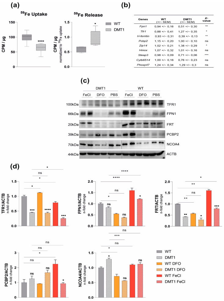
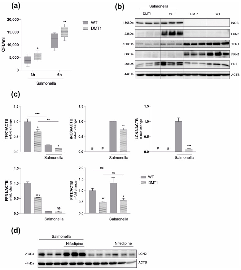
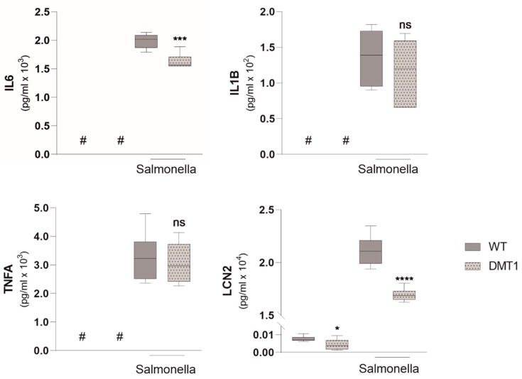
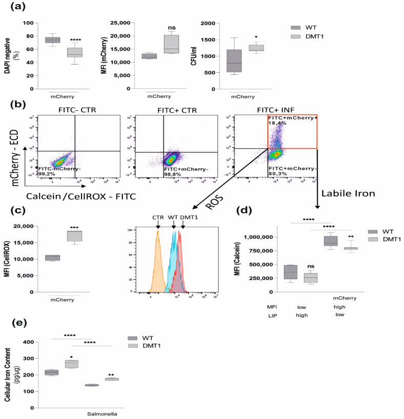
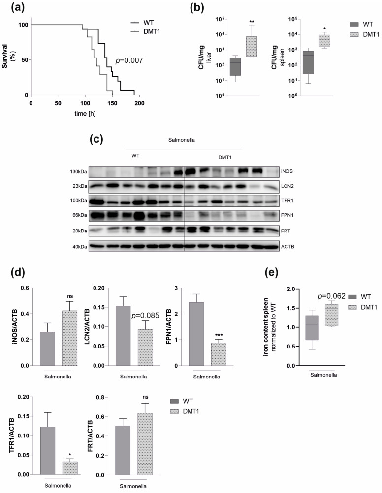
Macrophages are at the center of innate pathogen control and iron recycling. Divalent metal transporter 1 (DMT1) is essential for the uptake of non-transferrin-bound iron (NTBI) into macrophages and for the transfer of transferrin-bound iron from the endosome to the cytoplasm. As the control of cellular iron trafficking is central for the control of infection with siderophilic pathogens such as Salmonella Typhimurium, a Gram-negative bacterium residing within the phagosome of macrophages, we examined the potential role of DMT1 for infection control. Bone marrow derived macrophages lacking DMT1 (DMT1fl/flLysMCre(+)) present with reduced NTBI uptake and reduced levels of the iron storage protein ferritin, the iron exporter ferroportin and, surprisingly, of the iron uptake protein transferrin receptor. Further, DMT1-deficient macrophages have an impaired control of Salmonella Typhimurium infection, paralleled by reduced levels of the peptide lipocalin-2 (LCN2). LCN2 exerts anti-bacterial activity upon binding of microbial siderophores but also facilitates systemic and cellular hypoferremia. Remarkably, nifedipine, a pharmacological DMT1 activator, stimulates LCN2 expression in RAW264.7 macrophages, confirming its DMT1-dependent regulation. In addition, the absence of DMT1 increases the availability of iron for Salmonella upon infection and leads to increased bacterial proliferation and persistence within macrophages. Accordingly, mice harboring a macrophage-selective DMT1 disruption demonstrate reduced survival following Salmonella infection. This study highlights the importance of DMT1 in nutritional immunity and the significance of iron delivery for the control of infection with siderophilic bacteria.

Keywords: DMT1; Salmonella Typhimurium; iron; lipocalin 2; macrophage; nutritional immunity.


Conflict of interest statement

The authors declare no conflict of interest.

Figures

Figure 1
Figure 1
Role of DMT1 in bone-marrow-derived macrophages (BMDMs): (a) Uptake (n = 4) and release assay (n = 2): BMDMs were stimulated with radioactive 59Fe-citrate, and uptake/release was measured with a gamma-counter (counts per minute—cpm) and normalized to the protein concentration (µg). 59Fe release was normalized to uptake data; (b) Relative mRNA expression (n = 5) of genes involved in iron transport: Ribosomal Protein L4 (Rpl4) was used as a reference gene (averages ± SEM); (c) BMDMs (n = 2) were stimulated with 50 µM FerricChloride (FeCl) or Deferoxamine (DFO) overnight. Protein levels of transferrin receptor 1 (TFR1), ferroportin (FPN1), ferritin (FRT), Poly(rC)-binding protein 2 (PCBP2), Nuclear Receptor Coactivator 4 (NCOA4) and βActin(ACTB); (d) Densitometric quantification of Western blot results; see Figure S5 for a schematic overview of the discussed proteins. Data were compared by a two-tailed unpaired t-test (two groups) or analysis of variance (ANOVA) using the Bonferroni correction (more than two groups); n = mice per group; Data are expressed as box plots showing whiskers with minimum to maximum. Statistical significance: * p < 0.05, ** p < 0.01, *** p < 0.001, **** p < 0.0001, ns, no significance of differences; DMT1: DMT1 fl/flLysMCre(+), WT: wildtype.
Figure 2
Figure 2
DMT1 protects BMDMs against Salmonella infection. (a) Colony forming units (CFU) of Salmonella after 3 and 6 h respectively (n = 5); (b) Protein levels of inducible nitric oxide synthase (iNOS), lipocalin-2 (LCN2), transferrin receptor 1 (TFR1), ferroportin (FPN1), FRT (FRT) and βActin (ACTB) 6 h post infection (n = 3). (c) Densitometric quantification of Western blot results; (d) RAW264.7 macrophages were stimulated with 50 µM Nifedipine for 24 h. The protein level of LCN2 was determined via Western Blot analysis; Duplicates or triplicates from at least two independent experiments were compared by a two-tailed unpaired t-test (two groups) or analysis of variance (ANOVA) using a Bonferroni correction (more than two groups); n = mice per group. Error bars expressed as SEM. Box plots display whiskers with the minimum to maximum. Statistical significance * p < 0.05, ** p < 0.01, *** p < 0.001, ns, no significance of differences; #, below detection level; DMT1: DMT1 fl/flLysMCre(+), WT: wildtype.
Figure 3
Figure 3
DMT1 modulates Interleukin 6 and Lipocalin 2 formation. Enzyme linked immunosorbent assay (ELISA) from cell culture supernatants for Interleukin 6 (IL-6), Interleukin 1b (IL-1b), Tumor-necrosis factor alpha (TNFa), Lipocalin 2 (LCN2) (n = 2); Duplicates or triplicates from at least two independent experiments were compared by a two-tailed unpaired t-test (two groups) or analysis of variance (ANOVA) using Bonferroni correction (more than two groups); n = mice per group. Box plots display whiskers with the minimum to maximum. Statistical significance * p < 0.05, *** p < 0.001, **** p < 0.0001, ns, no significance of differences; #, below detection level; DMT1: DMT1 fl/flLysMCre(+), WT: wildtype.
Figure 4
Figure 4
DMT1 modulates iron availability in control and infected macrophages. Macrophages were infected with red fluorescent protein expressing Salmonella strains (mCherry) for 6 h. (a) The percentage of DAPI-negative cells indicating macrophage viability (n = 3) and mean fluorescence intensity (MFI) of mCherry infected macrophages (n = 3) suggesting mCherry proliferation. (b) Samples were stained either with CellROX Green (c) to determine oxidative stress or Calcein-AM (d) to measure LIP. A representative experiment with CellROX is shown using the following template: unstained control (CTR, FITC-mCherry−), stained control (FITC+mCherry−), stained infected (INF, FITC+mCherry+) BMDMs. (c) Reactive oxygen species (ROS) among mCherry (FITC+mCherry+)-containing macrophages were determined by flow cytometry. Data from 3 experiments are shown. Representative histogram including the FITC+mCherry-control normalized to mode is shown. (d) Calcein quench experiments were performed: DMT1 decreased the labile iron pool in infected macrophages (FITC+mCherry+) and uninfected macrophages (FITC+mCherry−), as evidenced by increasing Calcein-mediated mean fluorescence intensity (MFI). (e) Total iron content of control and infected macrophages was measured by atomic absorption (n = 2); Duplicates or triplicates from at least two independent experiments were compared by two-tailed unpaired t-test (two groups) or analysis of variance (ANOVA) using Bonferroni correction (more than two groups); n = mice per group. Box plots display whiskers with the minimum to maximum. Statistical significance * p < 0.05, ** p < 0.01, *** p < 0.001, **** p < 0.0001, ns, no significance of differences; DMT1: DMT1 fl/flLysMCre(+), WT: wildtype, mCherry: red fluorescent protein (RFP)-expressing Salmonella.
Figure 5
Figure 5
Macrophage DMT1 protects against Salmonella infection in vivo (a) Mice were subjected to a lethal dose of Salmonella by injecting 500 colony-forming units (CFU) intraperitoneally (n = 15). Survival data between control and mutant mice were compared using Cox regression and the Kaplan–Meier method using Wilcoxon’s test. (b) CFU normalized to the weight of spleen or liver from mice infected with Salmonella after 72 h (n = 7) (c) Western Blot for selected genes from spleens 72 h after infections (n = 7), (d) Densitometric quantification of the Western Blot, (e) Tissue iron of the spleen normalized to protein and wildtype control (n = 7). Duplicates or triplicates from at least two independent experiments were compared by a two-tailed unpaired t-test (two groups) or analysis of variance (ANOVA) using a Bonferroni correction (more than two groups); n = mice per group. Error bars express the SEM. Box plots display whiskers with the. minimum to maximum. Statistical significance * p < 0.05, ** p < 0.01, *** p < 0.001, ns, no significance of differences; DMT1: DMT1 fl/flLysMCre(+), WT: wildtype.

References

    1. Martins R., Carlos A.R., Braza F., Thompson J.A., Bastos-Amador P., Ramos S., Soares M.P. Disease Tolerance as an Inherent Component of Immunity. Annu. Rev. Immunol. 2019;37:405–437. doi: 10.1146/annurev-immunol-042718-041739. - DOI - PubMed
    1. Monteith A.J., Skaar E.P. The Impact of Metal Availability on Immune Function during Infection. Trends Endocrinol. Metab. 2021;32:916–928. doi: 10.1016/j.tem.2021.08.004. - DOI - PMC - PubMed
    1. Gerner R.R., Nuccio S.-P., Raffatellu M. Iron at the Host-Microbe Interface. Mol. Asp. Med. 2020;75:100895. doi: 10.1016/j.mam.2020.100895. - DOI - PMC - PubMed
    1. Oexle H., Gnaiger E., Weiss G. Iron-Dependent Changes in Cellular Energy Metabolism: Influence on Citric Acid Cycle and Oxidative Phosphorylation. Biochim. Biophys. Acta BBA-Bioenerg. 1999;1413:99–107. doi: 10.1016/S0005-2728(99)00088-2. - DOI - PubMed
    1. Muckenthaler M.U., Rivella S., Hentze M.W., Galy B. A Red Carpet for Iron Metabolism. Cell. 2017;168:344–361. doi: 10.1016/j.cell.2016.12.034. - DOI - PMC - PubMed

Substances