. 2022 Jun 20;23(12):6863.
doi: 10.3390/ijms23126863.

Roles of AGD2a in Plant Development and Microbial Interactions of Lotus japonicus

Affiliations

Roles of AGD2a in Plant Development and Microbial Interactions of Lotus japonicus

Mingchao Huang et al. Int J Mol Sci. .

Abstract

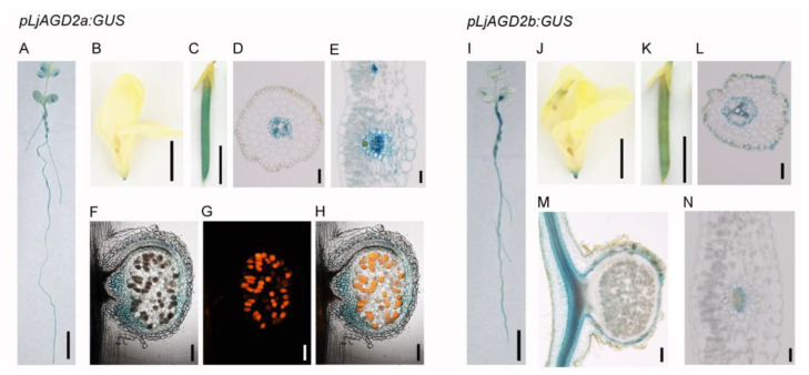
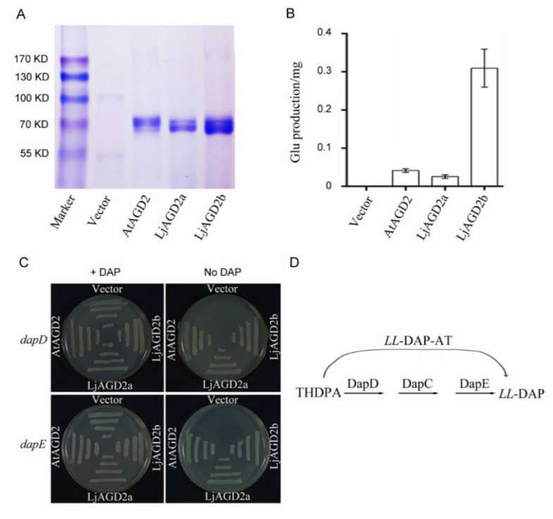
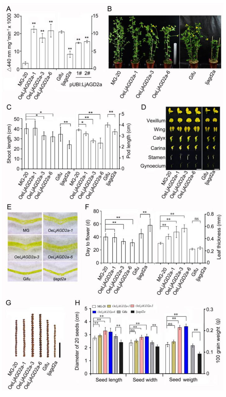
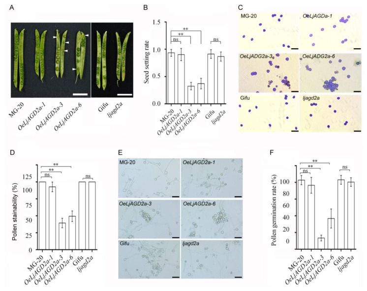
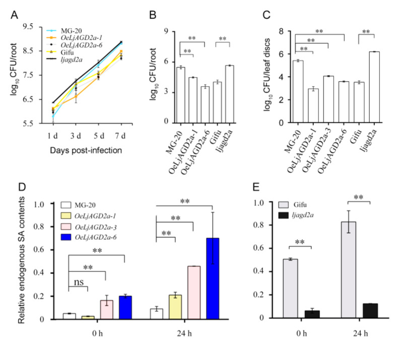
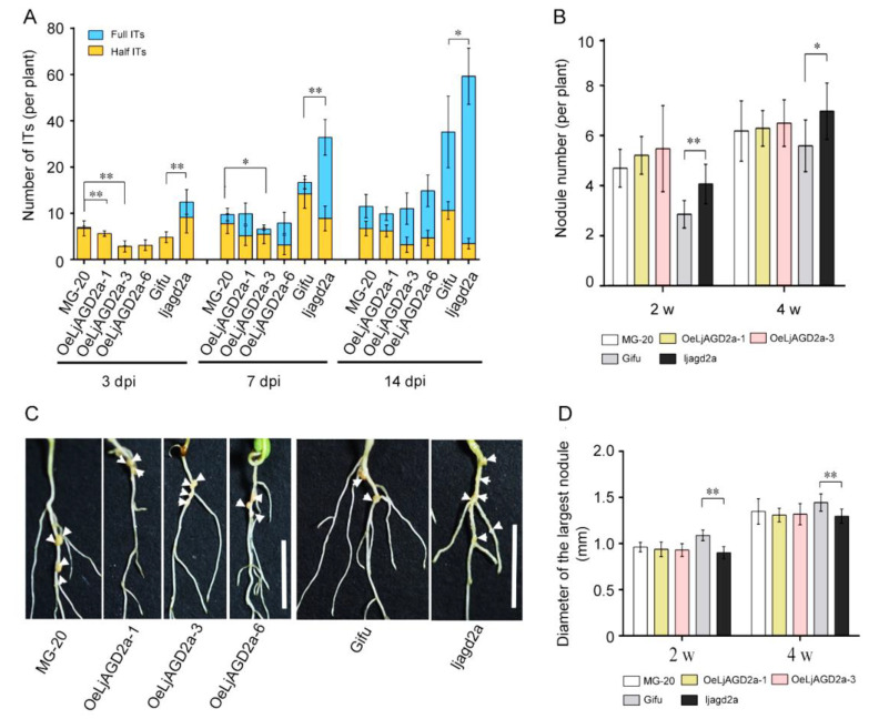
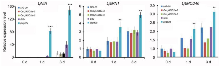
Arabidopsis AGD2 (Aberrant Growth and Death2) and its close homolog ALD1 (AGD2-like defense response protein 1) have divergent roles in plant defense. We previously reported that modulation of salicylic acid (SA) contents by ALD1 affects numbers of nodules produced by Lotus japonicus, but AGD2's role in leguminous plants remains unclear. A combination of enzymatic analysis and biological characterization of genetic materials was used to study the function of AGD2 (LjAGD2a and LjAGD2b) in L. japonicus. Both LjAGD2a and LjAGD2b could complement dapD and dapE mutants of Escherichia coli and had aminotransferase activity in vitro. ljagd2 plants, with insertional mutations of LjAGD2, had delayed flowering times and reduced seed weights. In contrast, overexpression of LjAGD2a in L. japonicus induced early flowering, with increases in seed and flower sizes, but reductions in pollen fertility and seed setting rates. Additionally, ljagd2a mutation resulted in increased expression of nodulin genes and corresponding increases in infection threads and nodule numbers following inoculation with Rhizobium. Changes in expression of LjAGD2a in L. japonicus also affected endogenous SA contents and hence resistance to pathogens. Our results indicate that LjAGD2a functions as an LL-DAP aminotransferase and plays important roles in plant development. Moreover, LjAGD2a activates defense signaling via the Lys synthesis pathway, thereby participating in legume-microbe interaction.

Keywords: Lotus japonicus; development; disease resistance; nodulation.


Conflict of interest statement

The authors declare no conflict of interest.

Figures

Figure 1
Figure 1
Expression pattern of LjAGD2s in L. japonicus. (AH) GUS histochemical staining of pLjAGD2a:GUS plants. (A) GUS histochemical staining of a 2-week-old pLjAGD2a:GUS seedling. (B) GUS staining of a flower of a pLjAGD2a:GUS plant. (C) GUS staining of a pod of a pLjAGD2a:GUS plant. (D) Section of a root of a 2-week-old pLjAGD2a:GUS plant. (E) Section of a leaf of a 2-month-old pLjAGD2a:GUS plant. (FH) Section of a nodule of a pLjAGD2a:GUS plant 2 weeks after inoculation with Mesorhizobium loti expressing DsRED. GUS histochemical staining of the nodule in (F), Bacteriods in the infection cells expressing red fluorescence in (G), Overlap of F and G images in (H). (IN) Histochemical staining of pLjAGD2b:GUS plants. (I) GUS histochemical staining of a 2-week-old pLjAGD2b:GUS seedling. (J) GUS staining of a flower of a pLjAGD2b:GUS plant. (K) GUS staining of a pod of a pLjAGD2b:GUS plant. (L) Section of a root of a 2-week-old pLjAGD2b:GUS plant. (M) Section of a nodule of a 2-week-old pLjAGD2b:GUS plant inoculated with M. loti. (N) Section of a leaf of a 2-month-old pLjAGD2b:GUS plant. Sixty seedlings were subjected to GUS staining as illustrated in (A,I), with more than 90% showing the phenotype. Flowers and pods collected from more than 10 individual plants were used for the GUS staining illustrated in (B,C,J,K). For sections, more than six samples were used.
Figure 2
Figure 2
Subcellular localization of LjAGD2a and LjAGD2b. LjAGD2a-eYFP and LjAGD2b-eYFP images indicating the localization of LjAGD2a and LjAGD2b in the chloroplasts of Arabidopsis protoplasts. The top row of images indicates the control. Scale bar = 10 µm.
Figure 3
Figure 3
Aminotransferase activities of AGD2s in E. coli. (A) Electrophoretic patterns of recombinant proteins purified using a Ni2+-nitrilotriacetic acid column. (B) Aminotransferase activities of AGD2s, assayed using 50 mM Lys and 50 mM 2-oxoglutarate as substrate and co-substrate, respectively. Aminotransferase activity was determined by measuring the concentration of the reaction product Glu. Bars indicate standard deviations (n = 3). (C) Complementation of dap mutants with AGD2. Strains AT980 (dapD mutant) and AT984 (dapE mutant) were transformed with either the plasmid vector (pGEX-KG) or an LjAGD2a, LjAGD2b and AtAGD2 (At4g33680) expression plasmid (pGEX-KG-AGD2). Colonies were selected on LB medium with 50 µg/mL DAP and 100 µg/mL ampicillin. Individual colonies were then plated onto NZY medium supplemented with 0.2% (w/v) Ara with or without 50 µg/mL DAP. The cultures were grown at 37 °C for 12 h. (D) Diagram of the DAP pathway in E. coli with the reaction catalyzed by LL-DAP-AT indicated.
Figure 4
Figure 4
Morphological effects of changing expression of LjAGD2a in L. japonicus. (A) LL-diaminopimelate aminotransferase (LL-DAP-AT) activities, showing a drastic reduction in ljagd2a and a significant increase in OeLjAGD2a lines (means with error bars indicating SD of five biological replicates). (B) Representative photograph of 8-week-old plants showing that OeLjAGD2a and ljagd2a plants had shorter roots than wild type plants (scale bar = 10 cm). (C) Shoot lengths, showing retarded growth of OeLjAGD2a and ljagd2a lines (Left). Both OeLjAGD2a and ljagd2a lines also produced shorter pods than wildtype plants (Right). (D) Photograph of 10-week-old plants showing that OeLjAGD2a plants had bigger flowers than wildtype plants, but insertional mutation of LjAGD2a did not change the flower size. Scale bar = 1 cm. (E) Sections showing that OeLjAGD2a plants had thick leaves. Scale bar = 1 mm. (F) OeLjAGD2a lines exhibited early flowering while insertional mutation of LjAGD2a delayed flowering (Left). Bar chart confirming that leaves of OeLjAGD2a lines were significantly thicker than wildtype leaves, but ljagd2a leaves did not significantly differ from wildtype leaves in thickness (Right). (G) Effects of changing LjAGD2a expression on seed morphology. Scale bar = 1 cm. (H) Seed sizes showing that LjAGD2a overexpression led to larger seeds, while ljagd2a seeds were smaller than wildtype seeds (Left). Measurement of 100-kernel weights of seeds showing that LjAGD2a overexpression and mutation resulted in increases and reductions in seed weight, respectively (Right). In the tests used to obtain results presented in (C,F,H), n = 20–30 per treatment. *, ** and ns indicate p < 0.05; p < 0.01 and no significant difference, respectively, according to the Duncan test. Error bars indicate the SD of three biological replicates.
Figure 5
Figure 5
Effects of changing expression of LjAGD2a on fertility in L. japonicus. (A) Immature seeds in OeLjAGD2a siliques. Aborted dead seeds are indicated by arrows. Scale bar = 1 cm. (B) Measured seed setting rates, which were dramatically decreased in OeLjAGD2a plants, relative to wildtype rates, but not significantly altered in ljagd2a mutants (means obtained from analyses of more than 50 siliques with standard deviations: ** indicate p < 0.01, respectively, according to the Duncan test). (C) Photograph showing morphology of pollen grains. (D) Stainability of pollen by iodine-potassium iodide solution, indicating viability. (E) Photograph showing results of in vitro pollen germination assays. (F) Pollen germination rates, based on analyses of pollen grains from 5 to 7 anthers of each of 8 to 10 plants (** and ns indicate p < 0.01 and no significant difference, respectively, according to the Duncan test). Scale bar = 50 µm in (C,D). Error bars in (B,D,F) indicate the SD of three biological replicates.
Figure 6
Figure 6
LjAGD2a promotes disease resistance in L japonicus. (A) Growth curves of R. solanacearum strains on the roots of transgenic ljagd2a lines, mutant lines and their respective wildtypes (MG-20 and Gifu129). Plants were infected with R. solanacearum at indicated time points. R. solanacearum grew more rapidly in ljagd2a plants than in their wildtype. In the first 3 days of pathogen infection, there was no significant difference in growth of R. solanacearum between OeLjAGD2a and MG-20 plants, but the pathogen’s growth was inhibited in OeLjAGD2a plants at 5 dpi. (B) Numbers of colony forming units (CFU) showing the disease susceptibility of roots. The indicated genotypes were inoculated with R. solanacearum by including the bacterium at a density equivalent to OD600 = 0.0002 in the growth medium and assaying the bacteria at 5 dpi. (C) Numbers of colony forming units (CFU) showing the disease susceptibility of detached leaves. Overexpression of LjAGD2a suppressed and its insertion mutation promoted growth of the pathogen. Detached leaves from 8-week-old plants were infected with R. solanacearum. (D) Total SA levels in OeLjAGD2a plants 0 and 24 h post-infection. (E) Total SA levels in ljagd2a mutants 0 and 24 h post-infection. Overexpression of LjAGD2a increased and its mutation reduced total SA levels after pathogen infection. 6–10 roots/leaves were used in each replicate and Error bars represent the SD of three replicates per genotype in (AC). In experiments providing results shown in (D,E) each sample had three to four replicates and error bars represent the SD of 3–4 replicates. In A–E, ** indicates p < 0.01 according to the Duncan test.
Figure 7
Figure 7
Changes in LjAGD2a expression affect L japonicus nodulation. (A) Numbers of infection threads (ITs) in transgenic plants and insertion mutants. Consistently higher numbers of ITs, relative to numbers in wildtype plants, were found in ljagd2a plants after M. loti inoculation. We also found fewer ITs in OeLjAGD2a plants at 3 days post-infection (dpi), but not 7 or 14 dpi. (B) Nodule numbers were higher in ljagd2a plants, but unchanged in OeLjAGD2a lines, relative to numbers in wildtype plants. (C) Nodules on roots 2 weeks after inoculation of plants. White arrows indicate nodules on the roots. (D) Measurements of the largest nodules on plants 4 weeks after inoculation with M. loti showing that mutation of LjAGD2a reduced nodule size. 15–20 plants were used in each replicate and three replicates per genotype were used in experiments providing data presented in A, B and D (±SD). * and ** indicate p < 0.5 and p < 0.01, respectively, according to the Duncan test.
Figure 8
Figure 8
Levels of transcript of nodulin genes—the nodule inception gene (LjNIN), ERF required for nodulation 1 (LjERN1) and early nodulin gene (LjENOD40)—in OeLjAGD2 plants and mutants. The expression of LjNIN, LjERN1 and LjENOD40 was increased in ljagd2a mutants after inoculation with M. loti, but overexpression of LjAGD2a did not change the expression of nodulin genes. The data were normalized using expression of Actin as an internal control. The Actin-normalized value for the 0-day wildtype sample was set to 1. Presented data are averages of triplicate RNA preparations (n = 15, ±SD). ** and *** indicate p < 0.01 and p < 0.001, respectively, according to the Duncan test.

Similar articles

Cited by

References

    1. Hartmann M., Zeier J. L-lysine metabolism to N-hydroxypipecolic acid: An integral immune-activating pathway in plants. Plant J. 2018;96:5–21. doi: 10.1111/tpj.14037. - DOI - PubMed
    1. Arruda P., Kemper E.L., Papes F., Leite A. Regulation of lysine catabolism in higher plants. Trends Plant Sci. 2000;5:324–330. doi: 10.1016/S1360-1385(00)01688-5. - DOI - PubMed
    1. Bunsupa S., Yamazaki M., Saito K. Quinolizidine alkaloid biosynthesis: Recent advances and future prospects. Front. Plant Sci. 2012;3:239. doi: 10.3389/fpls.2012.00239. - DOI - PMC - PubMed
    1. Arruda P., Barreto P. Lysine catabolism through the saccharopine pathway: Enzymes and intermediates involved in plant responses to abiotic and biotic stress. Front. Plant Sci. 2020;11:587. doi: 10.3389/fpls.2020.00587. - DOI - PMC - PubMed
    1. Hartmann M., Zeier T., Bernsdorff F., Reichel-Deland V., Kim D., Hohmann M., Scholten N., Schuck S., Bräutigam A., Hölze T., et al. Flavin monooxygenase-generated N-hydroxypipecolic acid is a critical element of plant systemic immunity. Cell. 2018;173:456.e16–469.e16. doi: 10.1016/j.cell.2018.02.049. - DOI - PubMed