. 2022 Jun 20;23(12):6870.
doi: 10.3390/ijms23126870.

In Vivo Inhibition of TRPC6 by SH045 Attenuates Renal Fibrosis in a New Zealand Obese (NZO) Mouse Model of Metabolic Syndrome

Affiliations

In Vivo Inhibition of TRPC6 by SH045 Attenuates Renal Fibrosis in a New Zealand Obese (NZO) Mouse Model of Metabolic Syndrome

Zhihuang Zheng et al. Int J Mol Sci. .

Abstract

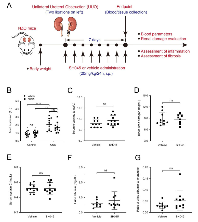
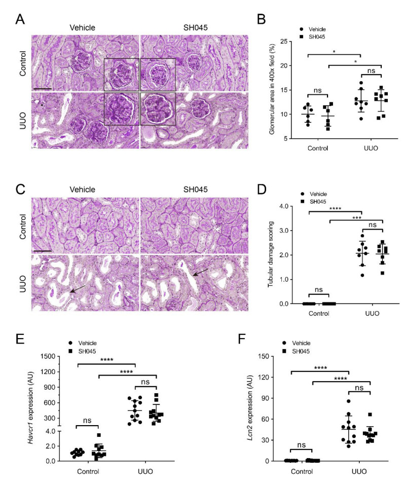
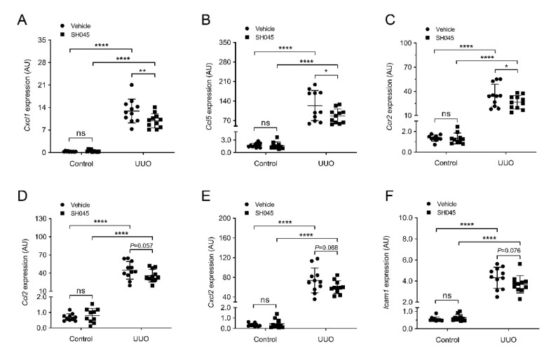
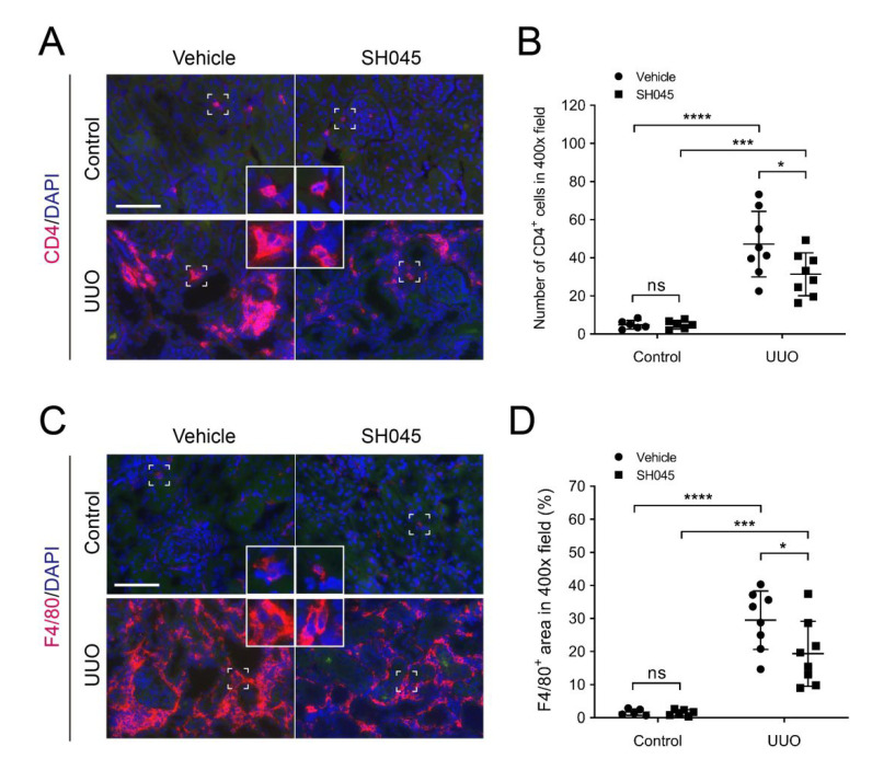
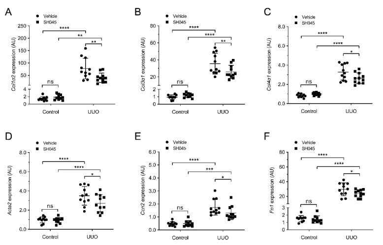
Metabolic syndrome is a significant worldwide public health challenge and is inextricably linked to adverse renal and cardiovascular outcomes. The inhibition of the transient receptor potential cation channel subfamily C member 6 (TRPC6) has been found to ameliorate renal outcomes in the unilateral ureteral obstruction (UUO) of accelerated renal fibrosis. Therefore, the pharmacological inhibition of TPRC6 could be a promising therapeutic intervention in the progressive tubulo-interstitial fibrosis in hypertension and metabolic syndrome. In the present study, we hypothesized that the novel selective TRPC6 inhibitor SH045 (larixyl N-methylcarbamate) ameliorates UUO-accelerated renal fibrosis in a New Zealand obese (NZO) mouse model, which is a polygenic model of metabolic syndrome. The in vivo inhibition of TRPC6 by SH045 markedly decreased the mRNA expression of pro-fibrotic markers (Col1α1, Col3α1, Col4α1, Acta2, Ccn2, Fn1) and chemokines (Cxcl1, Ccl5, Ccr2) in UUO kidneys of NZO mice compared to kidneys of vehicle-treated animals. Renal expressions of intercellular adhesion molecule 1 (ICAM-1) and α-smooth muscle actin (α-SMA) were diminished in SH045- versus vehicle-treated UUO mice. Furthermore, renal inflammatory cell infiltration (F4/80+ and CD4+) and tubulointerstitial fibrosis (Sirius red and fibronectin staining) were ameliorated in SH045-treated NZO mice. We conclude that the pharmacological inhibition of TRPC6 might be a promising antifibrotic therapeutic method to treat progressive tubulo-interstitial fibrosis in hypertension and metabolic syndrome.

Keywords: CKD; NZO mice; SH045; TRPC6; UUO; fibrosis; inflammation.


Conflict of interest statement

The authors declare no conflict of interest.

Figures

Figure 1
Figure 1
Impact of SH045 administration on renal function and Trpc6 expression in UUO model. (A) Experimental design of unilateral ureteral obstruction (UUO) model. NZO mice were subjected to UUO and then injected with SH045 (n = 11) or vehicle (n = 11) once every 24 h between day 0 and day 7. All mice were euthanized on day 7 after UUO surgery. (B) Renal mRNA levels of Trpc6 (control n = 10, UUO n = 11). Control group includes kidneys that were not subjected to the UUO. (C) Serum levels of creatinine, (D) blood urea nitrogen, and (E) cystatin C in the experimental UUO groups. (F) Urine albumin and (G) ratio of albumin to creatinine in the experimental UUO groups (UUO vehicle n = 10–11, UUO SH045 n = 8–11). Data expressed as means  ±  SD. Two-way ANOVA followed by Sidak’s multiple comparisons post hoc test. ** p < 0.01 and **** p < 0.0001 defined as significant. ns, not statistically significant. AU, arbitrary units.
Figure 2
Figure 2
SH045 impact on kidney histopathology after UUO. (A) Representative images of UUO-injured glomerulus (magnification: 400×). Kidney sections were stained with periodic acid–Schiff staining (PAS). (B) Quantification of glomerular damage (control n = 6, UUO n = 8). (C) Representative images of UUO-injured tubules (magnification: 400×). Kidneys sections were stained with periodic acid–Schiff staining (PAS). Arrows indicate tubular injury. Scale bars are 50 µm. (D) Semi-quantification of tubular damage (control n = 6, UUO n = 8). (E) Renal mRNA levels of kidney injury molecule 1 (Havcr1) and (F) Lipocalin 2 (Lcn2) (control n = 10, UUO n = 11). Data expressed as means ±  SD. Two-way ANOVA followed by Sidak’s multiple comparisons post hoc test. * p < 0.05, *** p < 0.001 and **** p < 0.0001 defined as significant. ns, not statistically significant. AU, arbitrary units.
Figure 3
Figure 3
SH045 impact on renal expression of inflammatory markers. (A) Renal mRNA levels of chemokine (C-X-C motif) ligand 1 (Cxcl1), (B) chemokine (C-C motif) ligand 5 (Ccl5), (C) chemokine (C-C motif) receptor 2 (Ccr2), (D) chemokine (C-C motif) ligand 2 (Ccl2), (E) chemokine (C-X-C motif) ligand 2 (Cxcl2), and (F) intercellular adhesion molecule-1 (Icam1) (control n = 10, UUO n = 11). Data expressed as means  ±  SD. Two-way ANOVA followed by Sidak’s multiple comparisons post hoc test. * p < 0.05, ** p < 0.01, and **** p < 0.0001 defined as significant. ns, not statistically significant. AU, arbitrary units.
Figure 4
Figure 4
SH045 impact on renal inflammatory cell accumulation after UUO. (A) Representative images of control and UUO-injured kidneys stained with CD4+ T cells (magnification: 400×). Rectangles represent single-cell magnifications. Scale bars are 50 µm. (B) Quantification in renal infiltration of CD4+ T cells (control n = 6, UUO n = 8). (C) Representative images of control and UUO-injured kidneys stained with F4/80+ macrophages (magnification: 400×). Rectangles represent single-cell magnifications. Scale bars are 50 µm. (D) Quantification in renal infiltration of F4/80+ macrophages (control n = 6, UUO n = 8). Data expressed as means  ±  SD. Two-way ANOVA followed by Sidak’s multiple comparisons post hoc test. * p < 0.05, *** p < 0.001, and **** p < 0.0001 defined as significant. ns, not statistically significant. AU, arbitrary units.
Figure 5
Figure 5
SH045 impact on expression of renal fibrotic markers UUO. (A) Renal mRNA levels of collagen type I α 1 (Col1α2), (B) Collagen type III α 1 (Col3α1), (C) Collagen type IV α 1 (Col4α1), (D) α-Smooth muscle actin (Acta2), (E) Connective tissue growth factor (Ccn2), and (F) Fibronectin (Fn1) (Control n = 10, UUO n = 11). Data expressed as means  ±  SD. Two-way ANOVA followed by Sidak’s multiple comparisons post hoc test. * p < 0.05, ** p < 0.01, *** p < 0.001 and **** p < 0.0001 defined as significant. ns, not statistically significant. AU, arbitrary units.
Figure 6
Figure 6
SH045 impact on renal fibrogenesis after UUO. (A) Representative images of control and UUO-injured kidneys stained with Sirius red (magnification: 400×). Scale bars are 50 µm. (B) Semi-quantification in renal Sirius red+ area proportion (control n = 6, UUO n = 8). (C) Representative images of control and UUO-injured kidneys stained with fibronectin (magnification: 400×). Scale bars are 50 µm. (D) Quantification in fibronectin+ area (control n = 6, UUO n = 8). (E) Representative images of control and UUO-injured kidneys stained with α-SMA (magnification: 400×). Scale bars are 50 µm. (F) Quantification of α-SMA+ staining (control n = 6, UUO n = 8). Data expressed as means  ±  SD. Two-way ANOVA followed by Sidak’s multiple comparisons post hoc test. * p < 0.05, ** p < 0.01, and **** p < 0.0001 defined as significant. ns, not statistically significant.

Similar articles

Cited by

References

    1. Jha V., Garcia-Garcia G., Iseki K., Li Z., Naicker S., Plattner B., Saran R., Wang A.Y., Yang C.W. Chronic kidney disease: Global dimension and perspectives. Lancet. 2013;382:260–272. doi: 10.1016/S0140-6736(13)60687-X. - DOI - PubMed
    1. Genovese G., Friedman D.J., Ross M.D., Lecordier L., Uzureau P., Freedman B.I., Bowden D.W., Langefeld C.D., Oleksyk T.K., Uscinski Knob A.L., et al. Association of trypanolytic ApoL1 variants with kidney disease in African Americans. Science. 2010;329:841–845. doi: 10.1126/science.1193032. - DOI - PMC - PubMed
    1. Li X., Pan J., Li H., Li G. DsbA-L mediated renal tubulointerstitial fibrosis in UUO mice. Nat. Commun. 2020;11:4467. doi: 10.1038/s41467-020-18304-z. - DOI - PMC - PubMed
    1. Black L., Lever J.M., Traylor A.M., Chen B., Yang Z., Esman S., Jiang Y., Cutter G., Boddu R., George J., et al. Divergent effects of AKI to CKD models on inflammation and fibrosis. Am. J. Physiol. Ren. Physiol. 2018;315:F1107–F1118. doi: 10.1152/ajprenal.00179.2018. - DOI - PMC - PubMed
    1. Schlondorff J. TRPC6 and kidney disease: Sclerosing more than just glomeruli? Kidney Int. 2017;91:773–775. doi: 10.1016/j.kint.2016.12.033. - DOI - PubMed

MeSH terms