. 2022 Jun 20;23(12):6875.
doi: 10.3390/ijms23126875.

Generation and Characterization of a Tumor Stromal Microenvironment and Analysis of Its Interplay with Breast Cancer Cells: An In Vitro Model to Study Breast Cancer-Associated Fibroblast Inactivation

Affiliations

Generation and Characterization of a Tumor Stromal Microenvironment and Analysis of Its Interplay with Breast Cancer Cells: An In Vitro Model to Study Breast Cancer-Associated Fibroblast Inactivation

Veronica Romano et al. Int J Mol Sci. .

Abstract

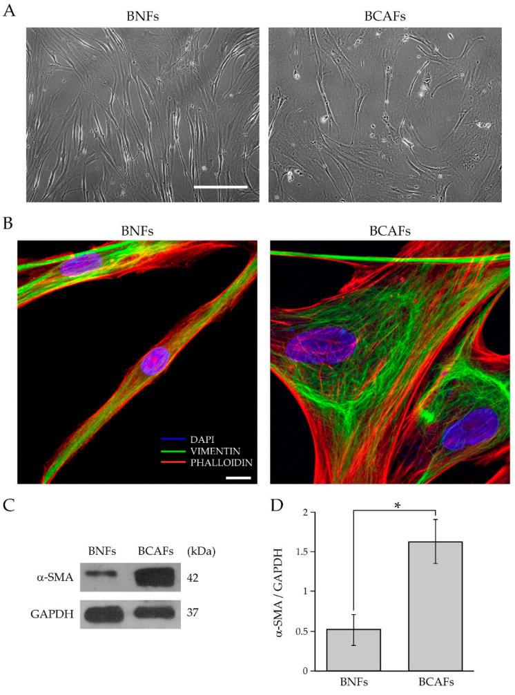
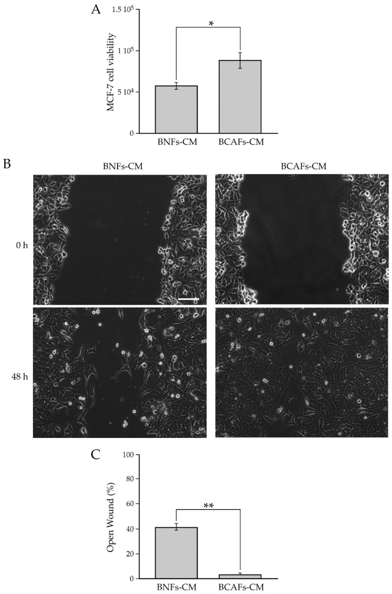
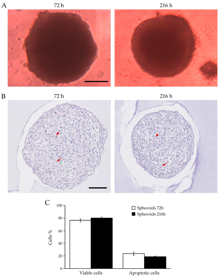
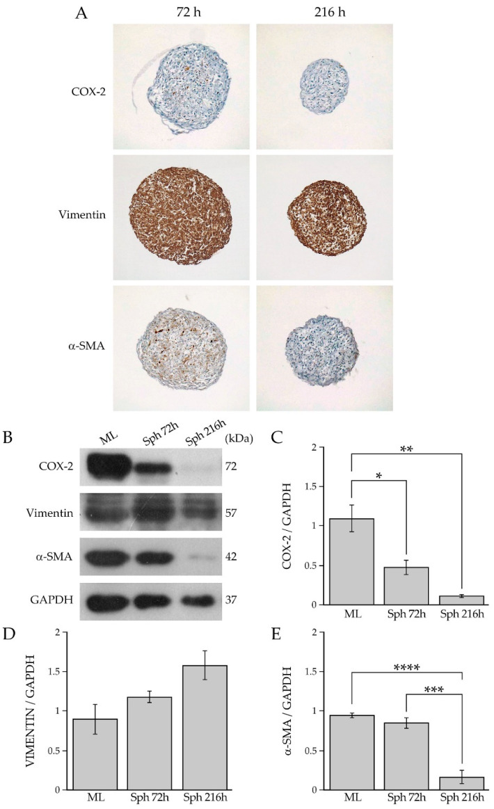
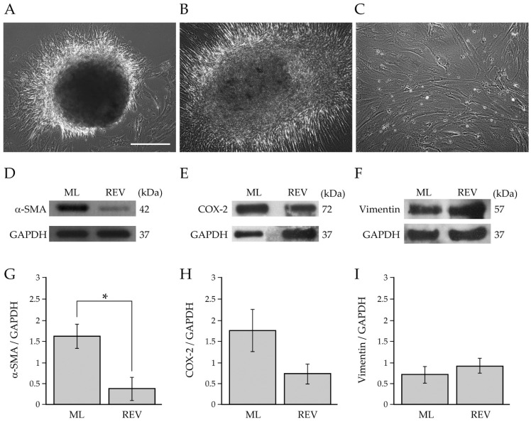
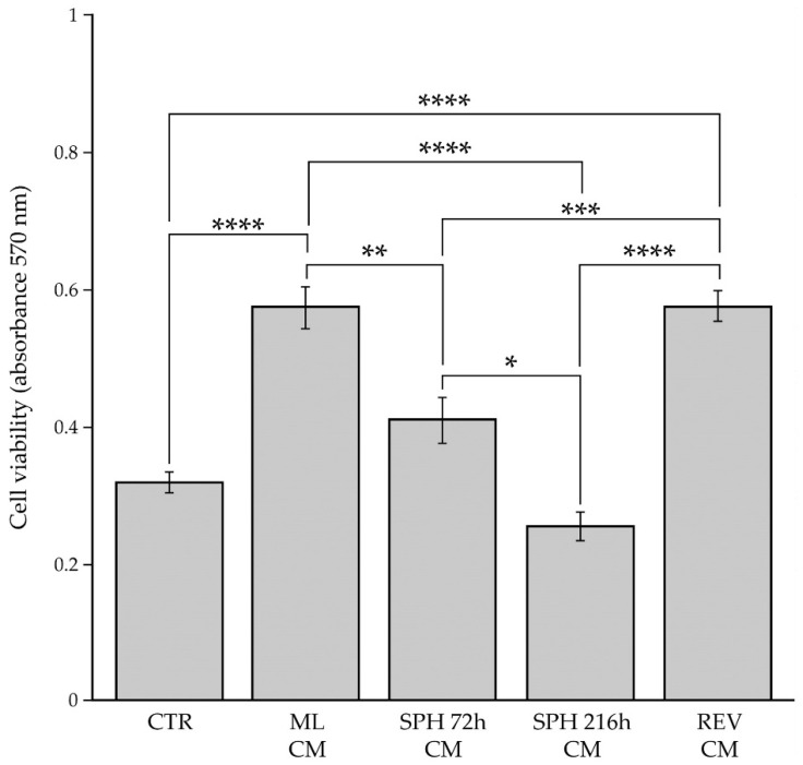
Breast cancer-associated fibroblasts (BCAFs), the most abundant non-cancer stromal cells of the breast tumor microenvironment (TME), dramatically sustain breast cancer (BC) progression by interacting with BC cells. BCAFs, as well as myofibroblasts, display an up regulation of activation and inflammation markers represented by α-smooth muscle actin (α-SMA) and cyclooxygenase 2 (COX-2). BCAF aggregates have been identified in the peripheral blood of metastatic BC patients. We generated an in vitro stromal model consisting of human primary BCAFs grown as monolayers or 3D cell aggregates, namely spheroids and reverted BCAFs, obtained from BCAF spheroids reverted to 2D cell adhesion growth after 216 h of 3D culture. We firstly evaluated the state of activation and inflammation and the mesenchymal status of the BCAF monolayers, BCAF spheroids and reverted BCAFs. Then, we analyzed the MCF-7 cell viability and migration following treatment with conditioned media from the different BCAF cultures. After 216 h of 3D culture, the BCAFs acquired an inactivated phenotype, associated with a significant reduction in α-SMA and COX-2 protein expression. The deactivation of the BCAF spheroids at 216 h was further confirmed by the cytostatic effect exerted by their conditioned medium on MCF-7 cells. Interestingly, the reverted BCAFs also retained a less activated phenotype as indicated by α-SMA protein expression reduction. Furthermore, the reverted BCAFs exhibited a reduced pro-tumor phenotype as indicated by the anti-migratory effect exerted by their conditioned medium on MCF-7 cells. The deactivation of BCAFs without drug treatment is possible and leads to a reduced capability of BCAFs to sustain BC progression in vitro. Consequently, this study could be a starting point to develop new therapeutic strategies targeting BCAFs and their interactions with cancer cells.

Keywords: aggregates; breast cancer; breast cancer cells; breast cancer-associated fibroblasts; conditioned medium; deactivation.


Conflict of interest statement

The authors declare no conflict of interest.

Figures

Figure 1
Figure 1
Characterization of fibroblasts isolated from normal and cancerous human breast tissues. (A) Representative cell morphology of BNFs and BCAFs by phase-contrast microscopy. Scale bar 200 µm. Magnification ×10. (B) Analysis of the cytoskeletal organization of BNFs and BCAFs by confocal fluorescence microscopy. Vimentin immunofluorescence (green channel) and phalloidin staining (red channel) of representative BNFs and BCAFs. DAPI (blue channel) was used to locate the nuclei. Images correspond to the 3D reconstruction of Z planes acquired from the top to the bottom of cells. Scale bar 10 µm. The images are representative of three independent experiments. (C) Western blotting analysis of α-SMA in protein extracts of BNFs and BCAFs; GAPDH was used as the loading control. A representative image of three independent experiments is shown. (D) Densitometric analyses of α-SMA protein levels. Data are reported as means of at least three independent experiments ± S.E.* p < 0.05.
Figure 2
Figure 2
(A) Influence of conditioned media from BNFs (BNFs-CM) and BCAFs (BCAFs-CM) on MCF-7 cell viability. Data are means of at least three independent experiments ± S.E. * p < 0.05. (B,C) Influence of conditioned media from BNFs (BNFs-CM) and BCAFs (BCAFs-CM) on MCF-7 cell migration. (B) Wound-Healing Assay performed on MCF-7 cells treated with fibroblast-conditioned media. Representative images of three independent experiments show the same fields with scratching at 0 and 48 h after wounding. Scale bar 100 µm. Magnification ×10. (C) Migratory capability quantification of MCF-7 cells. Wound widths were measured at 0 and 48 h after wounding. Data are expressed as percentage of fold decrease in open wound area compared with control (0 h) set as 100% and are reported as mean of three independent experiments ± S.E. ** p < 0.0001.
Figure 3
Figure 3
Real-Time Expression analysis of the CAF markers FAP and SPARC in human primary BCAFs (N = 3) and BC patient tissues (PT) (N = 3). Data are shown as percentage of mean +/− SD from triplicates of three independent experiments. Statistical analyses were conducted by one-way ANOVA with Tukey’s post hoc analysis. ns: not significant.
Figure 4
Figure 4
Analysis of BCAFs grown as spheroids. (A) Representative images of BCAF spheroids at 72 h and 216 h by phase-contrast microscopy. Scale bar 200 μm. Magnification ×10. Volumes of spheroids collected at 72 and 216 h were 0.109 mm3 ± 0.0075 and 0.0864 ± 0.0057, respectively. Data are reported as means of at least three independent experiments ± S.E. p < 0.05. (B) Hematoxylin and eosin staining of paraffin-embedded sections of BCAF spheroids collected at 72 and 216 h of 3D cell culture. Arrowheads indicate viable cells, while arrows indicate apoptotic cells. Scale bar 100 μm. Magnification ×20. (C) Live and apoptotic cell graph of spheroids collected at 72 h and 216 h.
Figure 5
Figure 5
(A) Immunohistochemical analysis of COX-2, vimentin and α-SMA in BCAF spheroids collected at 72 h and 26 h. Magnification ×10. (B) Western blotting analysis of COX-2, vimentin, and α-SMA in protein extracts of BCAF monolayers (ML) and BCAF spheroids collected at 72 h (SPH 72 h) and 216 h (SPH 216 h) of 3D cell culture on agar. GAPDH was used as the loading control. Representative images of three independent experiments are shown. Densitometric analyses of (C) COX-2, (D) vimentin and (E) α-SMA protein levels. Data are reported as means of three independent experiments ± S.E. * p < 0.05, ** p < 0.005, *** p < 0.001, **** p < 0.0005.
Figure 6
Figure 6
Characterization of reverted BCAFs. (A,B) Representative images of the outgrowth and spreading of reverted BCAFs from BCAF spheroids transferred on a conventional 2D cell culture dish to allow 2D cell growth and proliferation after 216 h of 3D cell culture on agar. Reverted BCAFs outgrowing from spheroid after (A) 1 day and (B) 1 week of 2D cell growth. (C) Representative cell morphology of reverted BCAFs by phase-contrast microscopy. Scale bar 200 µm. Magnification ×10. (DI) Western blotting analysis of α-SMA, COX-2 and vimentin in protein extracts of BCAF monolayers (ML) and reverted BCAFs (REV). GAPDH was used as loading control. Representative images of at least three independent experiments are shown. (GI) Densitometric analyses of α-SMA, COX-2 and vimentin protein levels. Data are reported as means of at least three independent experiments ± S.E. * p < 0.05.
Figure 7
Figure 7
Influence of tumor stromal microenvironment on the viability of BC cells. MCF-7 cells were grown for 48 h with the conditioned media derived from BCAF monolayers (ML CM), spheroids at 72 h (SPH 72 h CM), spheroids at 216 h (SPH 216 h CM) or reverted BCAFs (REV CM). Control is represented by MCF-7 cells incubated with unconditioned medium (CTR). The cell viability was evaluated by MTT assay. Data are means of three independent experiments ± S.E. * p < 0.005, ** p < 0.001, *** p < 0.0005, **** p < 0.0001.
Figure 8
Figure 8
Influence of BCAF monolayers and reverted BCAFs on the migratory capability of BC cells. (A) Wound-Healing Assay of MCF-7 cell line grown for 24 h with the conditioned media from BCAF monolayers (ML CM) and reverted BCAFs (REV CM). Standard cell culture medium was used as control. The representative images of three independent experiments show the same fields with scratching at 0 and 24 h after wounding. Scale bar 200 µm. Magnification ×10. (B) Migratory capability quantification of MCF-7 cells. Wound widths were measured at 0 and 24 h after wounding. Data are expressed as percentage of fold decrease in open wound area compared with control (0 h) set as 100% and are reported as mean of three independent experiments ± S.E. * p < 0.0005, ** p < 0.0001.

Similar articles

Cited by

References

    1. Ruocco M.R., Avagliano A., Granato G., Imparato V., Masone S., Masullo M., Nasso R., Montagnani S., Arcucci A. Involvement of Breast Cancer-Associated Fibroblasts in Tumor Development, Therapy Resistance and Evaluation of Potential Therapeutic Strategies. Curr. Med. Chem. 2018;25:3414–3434. doi: 10.2174/0929867325666180309120746. - DOI - PubMed
    1. Avagliano A., Fiume G., Ruocco M.R., Martucci N., Vecchio E., Insabato L., Russo D., Accurso A., Masone S., Montagnani S., et al. Influence of fibroblasts on mammary gland development, breast cancer microenvironment remodeling, and cancer cell dissemination. Cancers. 2020;12:1697. doi: 10.3390/cancers12061697. - DOI - PMC - PubMed
    1. Avagliano A., Ruocco M.R., Aliotta F., Belviso I., Accurso A., Masone S., Montagnani S., Arcucci A. Mitochondrial Flexibility of Breast Cancers: A Growth Advantage and a Therapeutic Opportunity. Cells. 2019;8:401. doi: 10.3390/cells8050401. - DOI - PMC - PubMed
    1. Li C.I., Anderson B.O., Daling J.R., Moe R.E. Trends in Incidence Rates of Invasive Lobular and Ductal Breast Carcinoma. J. Am. Med. Assoc. 2003;289:1421–1424. doi: 10.1001/jama.289.11.1421. - DOI - PubMed
    1. Makki J. Diversity of breast carcinoma: Histological subtypes and clinical relevance. Clin. Med. Insights Pathol. 2015;8:23–31. doi: 10.4137/CPath.S31563. - DOI - PMC - PubMed

Substances