

Decreased Expression of CIRP Induced by Therapeutic Hypothermia Correlates with Reduced Early Brain Injury after Subarachnoid Hemorrhage
- PMID: 35743480
- PMCID: PMC9225369
- DOI: 10.3390/jcm11123411
Decreased Expression of CIRP Induced by Therapeutic Hypothermia Correlates with Reduced Early Brain Injury after Subarachnoid Hemorrhage
Abstract
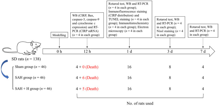
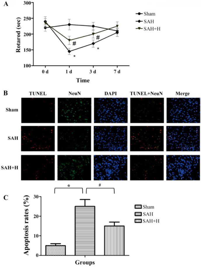
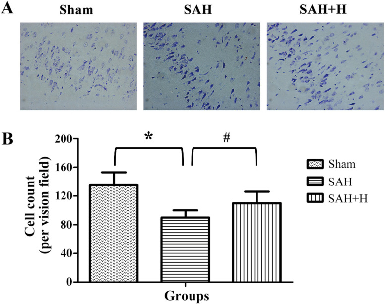
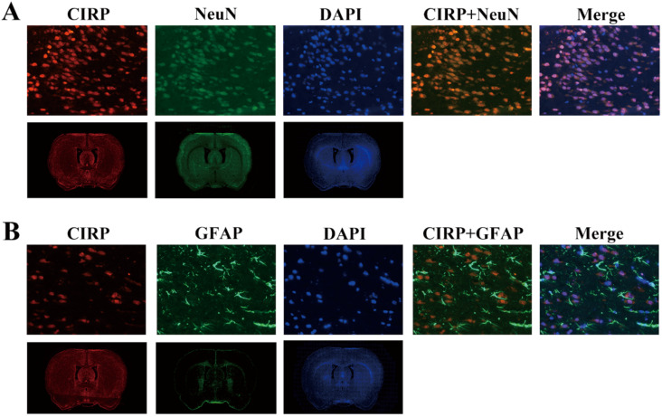
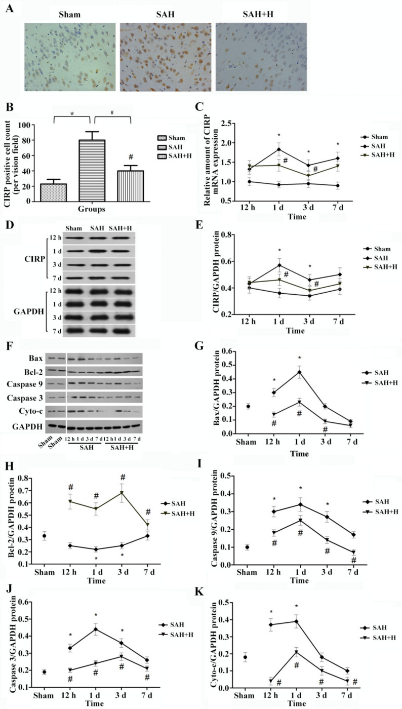
Early brain injury is considered to be a primary reason for the poor prognosis of patients suffering from subarachnoid hemorrhage (SAH). Due to its pro-inflammatory activity, cold-inducible RNA-binding protein (CIRP) has been implicated in the ischemic brain insult, but its possible interplay with hypothermia in SAH treatment remains to be evaluated. One-hundred and thirty-eight Sprague-Dawley rats (300-350 g males) were randomly allocated into the following groups: sham-operated (Sham); SAH; and SAH + hypothermia (SAH + H), each comprised of 46 animals. After treatments, the brain tissues of the three groups were randomly collected after 12 h, 1 d, 3 d, and 7 d, and the expression levels of the CIRP and mitochondrial apoptosis pathway-related proteins Bax, Bcl-2, caspase-9, caspase-3, and cytochrome c measured using Western blotting and real-time PCR. Brain damage was assessed by TUNEL and Nissl staining, the electron microscopy of brain tissue slices as well as functional rotarod tests. Expression of CIRP, Bax, caspase-9, caspase-3, and cytochrome c as well as reduced motor function incidence were higher in the SAH group, particularly during the first 3 d after SAH induction. Hypothermia blunted these SAH responses and apoptosis, thereby indicating reduced inflammatory signaling and less brain cell injury in the early period after SAH. Hypothermia treatment was found to effectively protect the brain tissue from early SAH injury in a rat model and its further evaluation as a therapeutic modality in SAH patients requires further study.
Keywords: apoptosis; cold-inducible RNA-binding protein (CIRP); early brain injury; hypothermia; subarachnoid hemorrhage (SAH).
Conflict of interest statement
The authors declare no conflict of interest.
Figures

Similar articles
-
Therapeutic Hypothermia Enhances Cold-Inducible RNA-Binding Protein Expression and Inhibits Mitochondrial Apoptosis in a Rat Model of Cardiac Arrest.Mol Neurobiol. 2017 May;54(4):2697-2705. doi: 10.1007/s12035-016-9813-6. Epub 2016 Mar 19. Mol Neurobiol. 2017. PMID: 26995407
-
Hydrogen-Rich Saline Attenuated Subarachnoid Hemorrhage-Induced Early Brain Injury in Rats by Suppressing Inflammatory Response: Possible Involvement of NF-κB Pathway and NLRP3 Inflammasome.Mol Neurobiol. 2016 Jul;53(5):3462-3476. doi: 10.1007/s12035-015-9242-y. Epub 2015 Jun 20. Mol Neurobiol. 2016. PMID: 26091790
-
GLP-1R Agonist Liraglutide Attenuates Inflammatory Reaction and Neuronal Apoptosis and Reduces Early Brain Injury After Subarachnoid Hemorrhage in Rats.Inflammation. 2021 Feb;44(1):397-406. doi: 10.1007/s10753-020-01344-4. Epub 2020 Sep 19. Inflammation. 2021. PMID: 32951103
-
Schisandrin B Inhibits NLRP3 Inflammasome Pathway and Attenuates Early Brain Injury in Rats of Subarachnoid Hemorrhage.Chin J Integr Med. 2022 Jul;28(7):594-602. doi: 10.1007/s11655-021-3348-z. Epub 2022 Jan 11. Chin J Integr Med. 2022. PMID: 35015222
-
Recombinant OX40 attenuates neuronal apoptosis through OX40-OX40L/PI3K/AKT signaling pathway following subarachnoid hemorrhage in rats.Exp Neurol. 2020 Apr;326:113179. doi: 10.1016/j.expneurol.2020.113179. Epub 2020 Jan 10. Exp Neurol. 2020. PMID: 31930990 Review.
Cited by
-
Changes in gene and protein expression related to feed intake and thermoregulation in broilers challenged with different doses of mixed Eimeria spp.Poult Sci. 2025 Jun 26;104(10):105481. doi: 10.1016/j.psj.2025.105481. Online ahead of print. Poult Sci. 2025. PMID: 40618565 Free PMC article.
References
-
- Bederson J.B., Connolly E.S., Jr., Batjer H.H., Dacey R.G., Dion J.E., Diringer M.N., Duldner J.E., Jr., Harbaugh R.E., Patel A.B., Rosenwasser R.H. Guidelines for the management of aneurysmal subarachnoid hemorrhage: A statement for healthcare professionals from a special writing group of the Stroke Council, American Heart Association. Stroke. 2009;40:994–1025. doi: 10.1161/STROKEAHA.108.191395. - DOI - PubMed
Grants and funding
LinkOut - more resources
Full Text Sources
Research Materials
