. 2022 May 25;12(6):785.
doi: 10.3390/life12060785.

The Emergence of Embryonic Myosin Heavy Chain during Branchiomeric Muscle Development

Affiliations

The Emergence of Embryonic Myosin Heavy Chain during Branchiomeric Muscle Development

Imadeldin Yahya et al. Life (Basel). .

Abstract

A prerequisite for discovering the properties and therapeutic potential of branchiomeric muscles is an understanding of their fate determination, pattering and differentiation. Although the expression of differentiation markers such as myosin heavy chain (MyHC) during trunk myogenesis has been more intensively studied, little is known about its expression in the developing branchiomeric muscle anlagen. To shed light on this, we traced the onset of MyHC expression in the facial and neck muscle anlagen by using the whole-mount in situ hybridization between embryonic days E9.5 and E15.5 in the mouse. Unlike trunk muscle, the facial and neck muscle anlagen express MyHC at late stages. Within the branchiomeric muscles, our results showed variation in the emergence of MyHC expression. MyHC was first detected in the first arch-derived muscle anlagen, while its expression in the second arch-derived muscle and non-somitic neck muscle began at a later time point. Additionally, we show that non-ectomesenchymal neural crest invasion of the second branchial arch is delayed compared with that of the first brachial arch in chicken embryos. Thus, our findings reflect the timing underlying branchiomeric muscle differentiation.

Keywords: MyHC; branchiomeric muscles; chicken embryos; mouse embryos; neck muscles; non-ectomesenchymal crest cells.


Conflict of interest statement

The authors declare that the research was conducted in the absence of any commercial or financial relationships that could be construed as a potential conflict of interest.

Figures
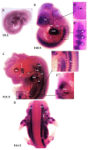
Figure 1
Figure 1
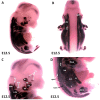
Expression of MyHC in early-stage mouse embryos. (AD) Whole-mount in situ hybridization for MyHC mRNA expression from E9.5–E11.5 of development. (B’,B”,C’,C”) higher magnifications of the areas indicated by the boxes in (B,C). (B) Note labeling of somites (white arrowheads), heart and first arch-derived muscles anlage by MyHC (black arrowhead). (C) MyHC is expressed in the first arch-derived muscle (te, ma) and second arch-derived muscle anlagen (bu). MyHC is also expressed in non-somitic neck muscles (a-trap, s-trap) and epaxial and hypaxial muscle anlagen. atp—acromiotrapezius; epm—epaxial musculature; flbm—fore limb muscle anlagen; hlbm—hind limb muscle anlagen; hpm—hypaxial muscle anlagen; mas—masseter; stp—spinotrapezius; te—temporal.
Figure 2
Figure 2
Expression of MyHC at stage E12.5. (A,B) Whole-mount in situ hybridization for MyHC. (C,D) Higher magnification of the photo in (A). (C) MyHC transcripts are detected strongly in the first arch-derived muscle anlagen (te, ma), neck muscles (a-trap, s-trap, stm) and second arch-derived muscle anlagen (bu, oo, zy). (D) MyHC transcripts are also observed in the thoracic and abdominal (ltd, icm, tr, ra), epaxial, hypaxial, fore limb and hind limb muscle anlagen. atp—acromiotrapezius; au—auricularis; epm—epaxial musculature; flbm—fore limb muscle anlagen; hlbm—hind limb muscle anlagen; hpm—hypaxial muscle anlagen; ltd—latissimus dorsi; mas—masseter; oo—orbicularis oculi; stp—spinotrapezius; stm—sternocleidomastoideus, te—temporalis, zy—zygomaticus.
Figure 3
Figure 3
Expression of MyHC at stage E13.5. (A) Scheme of facial and neck muscles. (B,C) Whole-mount in situ hybridization for MyHC. (D,E) Higher magnification of the photo in (A). Abbreviations as before and: sp—splenius. (CE) Similar to E12.5, MyHC labels the first and second arch-derived muscles, neck muscle, thoracic, abdominal and limb muscle anlagen. (D) MyHC expression is first seen at E13.5 in the splenius neck muscle anlagen.
Figure 4
Figure 4
Expression of MyHC at E14.5 and E15.5. (A,D) Whole-mount in situ hybridization for MyHC. (B,C) Higher magnification of the photo in (A). (E) Higher magnification of the photo in (D). (F) Scheme of facial muscles. Abbreviations as before and: oc—occipitalis. (C) MyHC is strongly expressed in the first and second arch-derived muscle as well as non-somitic neck muscle groups. (E) While second arch-derived muscle anlagen (oc, au, zy, fr, oo, qua and bu) show robust expression, MyHC transcripts are barely detectable in first arch-derived muscle anlagen (te, ma), neck, limbs, thoracic and abdominal muscle anlagen.
Figure 5
Figure 5
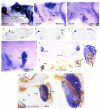
Relationship between non-ectomesenchymal crest and myogenic cells during development of chicken second branchial arch. (AC) Left lateral views of chicken embryos after whole mount in situ hybridization with Sox10 (black and red arrows denote hybridization signals in the first and second branchial arches). (A’C’) Vibratome frontal section of whole-mounted embryos in ((AC), indicated by the dotted line in (A)). (D) Whole-mount in situ hybridization for MyoG. (E) Vibratome frontal section of whole-mounted embryos in (D). (F,G) Immunostaining is performed for HNK1 on the same frontal section in (E) and sagittal section in (G) after whole-mount in situ hybridization. (F’,G’) Higher magnification of the area indicated by the box in (F). MyoG marked the myogenic cells in the second branchial arch (White arrow). The non-ectomesenchymal crest is labeled with HNK1 antibody; yellow arrows point to nerve. BA1—first branchial arch; BA2—second branchial arch; ov—otic vesicle; V—trigeminal ganglion; VII—facial ganglion.

Similar articles

Cited by

References

    1. Nogueira J.M., Hawrot K., Sharpe C., Noble A., Wood W.M., Jorge E.C., Goldhamer D.J., Kardon G., Dietrich S. The emergence of Pax7-expressing muscle stem cells during vertebrate head muscle development. Front. Aging Neurosci. 2015;7:62. doi: 10.3389/fnagi.2015.00062. - DOI - PMC - PubMed
    1. Noden D.M., Francis-West P. The differentiation and morphogenesis of craniofacial muscles. Dev. Dyn. 2006;235:1194–1218. doi: 10.1002/dvdy.20697. - DOI - PubMed
    1. Yahya I., Morosan-Puopolo G., Brand-Saberi B. The CXCR4/SDF-1 Axis in the Development of Facial Expression and Non-somitic Neck Muscles. Front. Cell Dev. Biol. 2020;8:615264. doi: 10.3389/fcell.2020.615264. - DOI - PMC - PubMed
    1. Buckingham M., Relaix F. PAX3 and PAX7 as upstream regulators of myogenesis. Semin. Cell Dev. Biol. 2015;44:115–125. doi: 10.1016/j.semcdb.2015.09.017. - DOI - PubMed
    1. Sambasivan R., Gayraud-Morel B., Dumas G., Cimper C., Paisant S., Kelly R.G., Tajbakhsh S. Distinct regulatory cascades govern extraocular and pharyngeal arch muscle progenitor cell fates. Dev. Cell. 2009;16:810–821. doi: 10.1016/j.devcel.2009.05.008. - DOI - PubMed

LinkOut - more resources