Review
. 2022 May 29;12(6):809.
doi: 10.3390/life12060809.

Fetal Brain Development: Regulating Processes and Related Malformations

Affiliations
Review

Fetal Brain Development: Regulating Processes and Related Malformations

Zvi Leibovitz et al. Life (Basel). .

Abstract

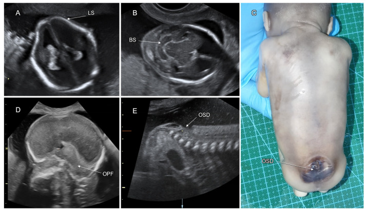
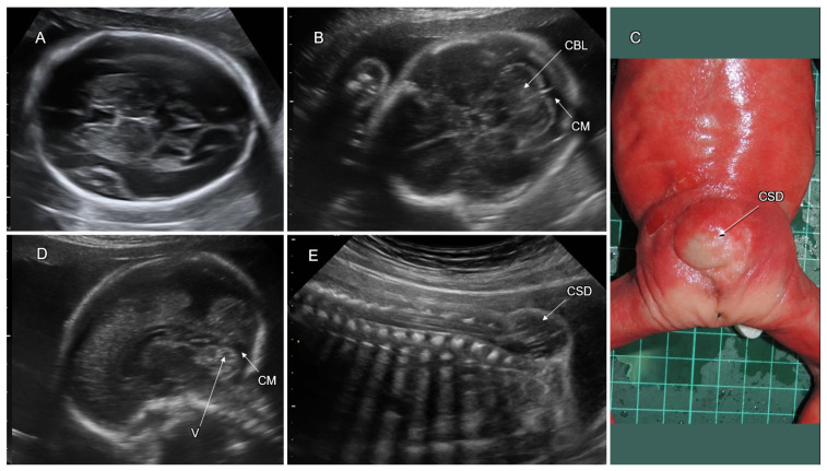
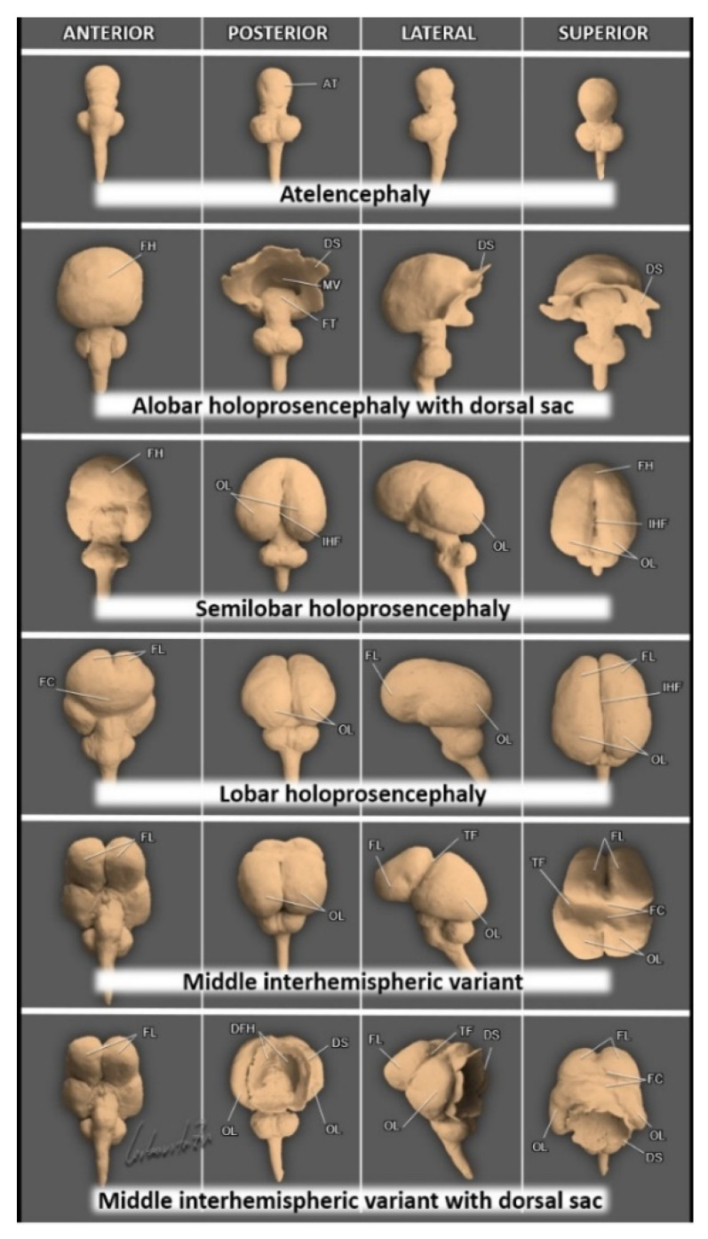
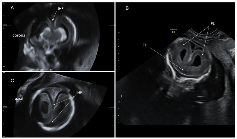
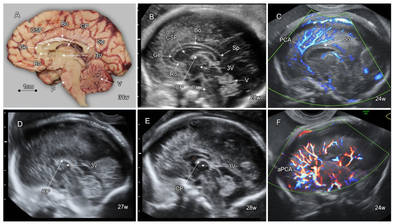
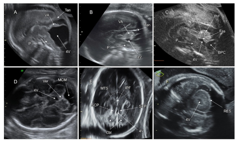
This paper describes the contemporary state of knowledge regarding processes that regulate normal development of the embryonic-fetal central nervous system (CNS). The processes are described according to the developmental timetable: dorsal induction, ventral induction, neurogenesis, neuronal migration, post-migration neuronal development, and cortical organization. We review the current literature on CNS malformations associated with these regulating processes. We specifically address neural tube defects, holoprosencephaly, malformations of cortical development (including microcephaly, megalencephaly, lissencephaly, cobblestone malformations, gray matter heterotopia, and polymicrogyria), disorders of the corpus callosum, and posterior fossa malformations. Fetal ventriculomegaly, which frequently accompanies these disorders, is also reviewed. Each malformation is described with reference to the etiology, genetic causes, prenatal sonographic imaging, associated anomalies, differential diagnosis, complimentary diagnostic studies, clinical interventions, neurodevelopmental outcome, and life quality.

Keywords: CNS malformations; brain development; fetal neurology.


Conflict of interest statement

The authors declare no conflict of interest.

Figures

Figure 1
Figure 1
Formation of the neural tube. Legend: (A) An upper view of the embryonic disk at the 3rd postconceptional week. ED, the ectodermal surface of the embryonic disk; NF, neural folds; FNF, initial fusion of the neural folds, forming a neural tube (NT); PS, primitive streak. (B) A mid-sagittal section of a human embryo depicting closure sites (CS 1–5) of the neural tube (NT). Types of neural tube defects related to failed fusion of the neural folds at the specific closure sites (CS 1–5) are indicated in the text boxes. NT, neural tube; SD, spinal dysraphism; SN, secondary neurulation (the formation of the most distal part of the neural tube from the multipotential stem cells in the tail bud by longitudinal growth and canalization, indicated by the dashed arrow).
Figure 2
Figure 2
Models of early neural tube development. Legend: At the 3-vesicle stage, by the end of the fourth postconceptional week, three primitive brain vesicles are formed along the rostral part of the neural tube: the prosencephalon (Pro), mesencephalon (Mes), and rhombencephalon (Rho). At the 5-vesicle stage, by the seventh postconceptional week, the cephalic part of the prosencephalon rapidly grows and divides laterally into the two telencephalic vesicles (Tel) and a distinct medial diencephalic vesicle (Die). The rhombencephalon is divided into the metencephalon (Met) and the myelencephalon (Mye).
Figure 3
Figure 3
Progression from acrania to anencephaly. Legend: (A) A three-dimensional sonographic multiplanar reconstruction image demonstrating the coronal, mid-sagittal, and axial head planes of a fetus with acrania at 11 gestational weeks. Note the absent cranial vault and dysmorphic cerebral hemispheres (HS) floating in amniotic fluid above the base of the skull (exencephaly). (B,C) Three-dimensional sonographic surface render images of the fetal head at 27 gestational weeks. Note the flat, irregular area cerebrovasculosa (AC), a neural-vascular tissue remaining after traumatic destruction of the exposed cerebral hemispheres, the absent forehead, and protruding eyes. (D,E) Postpartum photographs corresponding to (B,C). The baby died 10 days after birth.
Figure 4
Figure 4
Fetal posterior cephaloceles at the late first and early second trimesters. Legend: (A,B) Three-dimensional sonographic multiplanar reconstruction images demonstrating the coronal, mid-sagittal, and axial head planes of two fetuses with posterior cephalocele. (A) An occipitocervical meningocele (M) without neural tissue inside the lesion at 12 gestational weeks. (B) Occipital encephalocele (EN), containing herniated tectum and cerebellum (TC) at 17gestational weeks; note the dilated lateral ventricles (VM).
Figure 5
Figure 5
Huge occipital encephalocele. Legend: (A,C) Mid-sagittal and axial head sonograms of a fetus at 24 gestational weeks with a huge occipital encephalocele (EN) containing herniated occipital lobes (OL) and dysmorphic tectum and cerebellum (TC). (B) Postpartum photograph of the lesion; note the associated microcephaly. The baby died 3 days after birth.
Figure 6
Figure 6
Rare forms of cephalocele. Legend: (A,B) Mid-sagittal and axial head sonograms of a fetus at 19 gestational weeks with a frontal encephalocele (EN) protruding through the cranial defect between the nasal and frontal bones. (C) Mid-sagittal sonograms of a fetus at 18 gestational weeks demonstrating a huge occipital encephalocele (EN); almost all the brain is located outside the calvarium connected to the amniotic band (AB); (D) Lateral photograph of the abortus (the exposed brain tissue was destroyed during abortion); note the amniotic bands (AB) connected to the cranial defect (EN) and associated malformations: the total absence of the left upper limb (L), omphalocele (O), and bilateral clubfoot (CL).
Figure 7
Figure 7
Sonographic features of open spinal dysraphism in a fetus at 20 gestational weeks. Legend: (A) Axial transventricular head plane, note the lemon-shaped fetal head (LS) due to scalloping of the frontal bones (the lemon sign). (B) Axial transcerebellar head plane, note the banana sign (BS): the curved cerebellar shape caused by the posterior displacement of the small brain towards the cranial vault within the obliterated cisterna magna. (D) Mid-sagittal head plane, note the small obliterated posterior fossa (OPF) with downward displacement of the brainstem, cerebellar tonsils, and vermis into the foramen magnum (Chiari-II malformation). (E) Mid-sagittal section through the lower spine. Note the open lumbosacral spinal dysraphism (OSD). (C) Posterior photograph of the abortus. Note that the lesion (OSD) is covered by a thin translucent membrane without normal skin.
Figure 8
Figure 8
Sonographic features of closed spinal dysraphism in a fetus at 25 gestational weeks Legend: (A) Axial transthalamic head plane; note the normal shape of the fetal head. (B) Axial transcerebellar head plane; note the normal cerebellum (CBL) and cisterna magna (CM). (D) Mid-sagittal head plane; note the normal anatomy of the posterior fossa with a well-formed vermis (V) and a normal cisterna magna (CM). (E) Mid-sagittal section through the lower spine. Note the closed sacral spinal dysraphism (CSD) containing neural tissue (myelomeningocele). (C) Posterior photograph of the abortus. Note that the lesion (CSD) presents as a subcutaneous mass covered by normal skin.
Figure 9
Figure 9
Models of the holoprosencephaly spectrum. Legend: Atelencephaly: rudimentally formed fused telencephalons (AT). Alobar holoprosencephaly: completely fused hemispheres (FH) lacking any separation with a single undivided midline ventricle (MV). Note the fused thalami (FT) and the ruptured dorsal sac (DS). Semilobar holoprosencephaly: the cerebral hemispheres are fused anteriorly (FH); the occipital lobes (OL) are separated by the interhemispheric fissure (IHF). Lobar holoprosencephaly: the cerebral hemispheres are almost completely separated. Fusion of the hemispheres occurs only in the rostral-inferior frontal cortex (FC). FL, divided frontal lobes above the fused cortex. Middle interhemispheric variant (MIH): the hemispheric fusion (FC) takes place only between the posterior frontal and parietal lobes. Note the abnormal transverse fissure across the midline (TF). Middle interhemispheric variant (MIH) with dorsal sac: the hemispheres are fused (FC) only between the posterior frontal and parietal lobes. Note the abnormal transverse fissure across the midline (TF), the divided frontal horns (DFN) of the lateral ventricles, and the ruptured dorsal sac (DS) separating small occipital lobes (OL).
Figure 10
Figure 10
Sonographic features of alobar holoprosencephaly in a fetus at 18 gestational weeks. Legend: (A) A three-dimensional sonographic multiplanar reconstruction image demonstrating the coronal, mid-sagittal, and axial head planes. Note complete fusion of the hemispheres (FH), the undivided monoventricle (MV), and the thalamic fusion (FT), resulting in obstruction of the CSF flow into the aqueduct of Sylvius and the formation of a large dorsal cyst (DS). (B,C) Posterior photographs of the autopsy brain specimens, demonstrating the monoventricle, fused hemispheres (FH) and thalami (FT); note the ruptured membranes of the dorsal sac (DS), see also Figure 9.
Figure 11
Figure 11
Sonographic features of lobar holoprosencephaly in a fetus at 19 gestational weeks. Legend: (A,C) Axial and coronal head planes. The cerebral hemispheres are separated along the upper aspect of the interhemispheric fissure (IHF). (B) Coronal transfrontal head plane. Fusion of the hemispheres occurs only in the rostral-inferior frontal cortex (FC). The frontal lobes (FL) are divided above the fused cortex. Note complete fusion of the hemispheres (FH), the undivided monoventricle (MV), and the thalamic fusion (FT), resulting in obstruction of the CSF flow into the aqueduct of Sylvius and the formation of a large dorsal cyst (DS). (B,C) Posterior photographs of the autopsy brain specimens, demonstrating the monoventricle, fused hemispheres (FH) and thalami (FT); note the ruptured membranes of the dorsal sac (DS), see also Figure 9.
Figure 12
Figure 12
Fetal corpus callosum in the mid-sagittal plane: normal structure and callosal agenesis. Legend: (A) Median photograph of the brain specimen at 34 weeks, demonstrating the normal structural segments of the corpus callosum: the rostrum (Ro), genu (Ge), body (Bo), and splenium (Sp). Note the cavum septi pellucidi (CSP) below the corpus. 3V, the third ventricle; P, the pons; and V, the vermis. (BE) Mid-sagittal sonographic fetal head planes: (B) A normal fetus at 28 weeks of gestation. Note the anatomical structures indicated in panel A. CP, the choroid plexus of the third ventricle. (C) A normal fetus at 24 gestational weeks. The regular path of the pericallosal artery (PCA), applying high-definition Doppler, is shown. (D) A fetus at 27 gestational weeks with complete agenesis of the corpus callosum. (E) A fetus at 28 gestational weeks with partial agenesis of the corpus callosum; note the lack of the rostrum, genu, and splenium. (F) A fetus with complete agenesis of the corpus callosum at 24 gestational weeks. Note an abnormal branching of the pericallosal artery (aPCA) demonstrated by high-definition Doppler.
Figure 13
Figure 13
CNS imaging features of abnormal callosal development. Legend: (A,B) Transthalamic coronal planes: (A) A normal fetus at 24 gestational weeks. Note the normal oblique alignment of the anterior horns (AH) relative to the corpus callosum (CC), appearing as a concave hypoechoic stripe above the cavum septi pellucidi (CSP). (B) A fetus with complete callosal agenesis at 22 gestational weeks. Note the vertically oriented and remote anterior horns, the absence of the CSP, and the wide interhemispheric fissure (IHF). (C,D) Axial transventricular head planes of a fetus with complete callosal agenesis at 27 gestational weeks. Note the colpocephalic configuration of the lateral ventricle, the “tear drop” sign (TD), remote and parallelly oriented anterior horns (AH), ventriculomegaly, and wide interhemispheric fissure (IHF). (E) Sagittal head plane of a fetus with complete callosal agenesis (pACC) at 39 gestational weeks. Note the abnormal radial sulci (RS) converging towards the 3rd ventricle and a lack of the cingulate sulcus (the “sunray” sign). (F) Median photograph of the brain specimen at 34 weeks. Note the abnormal radial sulci (RS) converging towards the 3rd ventricle and a lack of the cingulate sulcus (the “sunray” sign).
Figure 14
Figure 14
Fetal sonographic features of malformations of cortical development. Legend: (A,B) Transthalamic coronal and axal planes of a fetus with lissencephaly at 24 gestational weeks. Note the shallow Sylvian fissures (SF). (C,D) Transventricular axial and sagittal planes of a fetus with Walker–Warburg syndrome at 26 gestational weeks. Note the flat Sylvian fissures (SF), severe dilatation of the lateral (VM) and third (3V) ventricles, and the typical Z-shaped hypoplastic brainstem (BS). (E,F) Transventricular axial and sagittal ventricular planes of a female fetus with periventricular nodular heterotopia at 32 gestational weeks. Note the chain of the small subependymal protrusions bulging into the ventricle (PNH). (G) Transthalamic coronal plane of a fetus with bilateral frontal polymicrogyria at 25 gestational weeks. Note an irregular, serrated cortical contour (PMG). (H) Transthalamic coronal planes of a fetus with schizencephaly at 22 gestational weeks. Note the extensive lateral parenchymal cleft (SCH) extending from the cerebral cortex to the ventricle associated with the absence of the cavim septi pellucidi.
Figure 15
Figure 15
Posterior fossa malformations. Legends: (AC) Mid-sagittal head planes: (A) A fetus at 22 weeks of gestation with Dandy–Walker malformation. Note the enlarged posterior fossa with upward displacement of the tentorium cerebelli (Ten), cystic dilatation of the 4th ventricle (4V) that fills the entire posterior fossa, and almost complete agenesis and substantial upward rotation of the vermis (VA). (B) A fetus at 28 gestational weeks with inferior vermian agenesis. Note the small vermis with mild upward rotation, lacking all the inferior lobules (VA). The tentorium (Ten) is in a normal position. The fastigium and the primary vermian fissure are not formed (compared with a normal vermian structure in panel (C). (C) A fetus at 25 gestational weeks with a Blake’s pouch cyst (BPC). Note the wall of the cyst, protruding from the floor of the fourth ventricle into the cisterna magna. The vermis is of normal size and shape and only slightly rotated upwards. Note the normal fastigial beak (Fas) in the middle of the intraventricular vermian contour and the normally located primary vermian fissure (PrF) dividing between the upper and lower vermian regions in a 1:2 ratio. (DF) Axial transcerebellar head planes: (D) A fetus at 27 gestational weeks with megacisterna magna (MCM). Note the vermian tissue that completely separates between the normally shaped 4th ventricle (4V) and the symmetrically enlarged cisterna magna (MCM). (E) Axial plane through the upper cerebellar region of a fetus at 25 gestational weeks with Joubert syndrome. Note the absence of the vermis and the elongated shape of the 4th ventricle (4V) (compared with normal anatomy in panel (C). Note the “molar tooth sign” (MTS) encircled by a dashed ellipse, depicting the typically elongated and thickened superior cerebellar peduncles (SCP) running on both sides of the 4th ventricle (4V) and the deep interpeduncular fossa (IPF). (F) A fetus at 19 gestational weeks with rhombencephalosynapsis. Note median fusion of the cerebellar hemispheres (Cbl), resulting in a small and round-shaped cerebellum lacking the vermian tissue and the posterior cerebellar notch (RES).
Figure 16
Figure 16
Fetal ventriculomegaly and associated disorders. Legend: (AF) Fetal head planes on axial sonography: (A) A fetus at 31 weeks of gestation with global severe ventriculomegaly (VM) due to aqueductal stenosis. Note the thin and regular ependymal lining; the third ventricle is dilated (not shown). (B) A fetus at 29 gestational weeks with complete agenesis of the corpus callosum. Note the colpocephalic dilatation of the atrium and occipital horn (VM). (C) A fetus at 26 gestational weeks with a huge intracranial teratoma (Tu). Note a prominent mass effect resulting in midline displacement and severe contralateral ventriculomegaly (VM). (D) A fetus at 36 gestational weeks with intraventricular hemorrhage grade-3. Note a thick and irregular ependymal lining, dilated third ventricle, and intraventricular blood clots (BC). (E) A fetus at 34 gestational weeks with a vein of Galen aneurysmal malformation (VGAM). Note severe bilateral ventriculomegaly (VM) due to the mass effect of the VGAM, leading to obstruction of the CSF flow from the third ventricle. (F) A fetus at 36 gestational weeks with intrauterine CMV infection. Note bilateral ventriculomegaly (VM) and diffuse periventricular calcifications (Calc).

References

    1. Paladini D., Malinger G., Timor-Tritsch I.E. ISUOG Practice Guidelines (updated), sonographic examination of the fetal central nervous system. Part 2, performance of targeted neurosonography. Ultrasound Obstet. Gynecol. 2021;57:661–671. doi: 10.1002/uog.23616. - DOI - PubMed
    1. Govaert P., Triulzi F. Handbook of Clinical Neurology. Volume 171. Elsevier; Amsterdam, The Netherlands: 2020. The developing brain by trimester; pp. 245–289. - DOI - PubMed
    1. Silbereis J.C., Pochareddy S. The Cellular and Molecular Landscapes of the Developing Human Central Nervous System. Neuron. 2016;89:248–268. doi: 10.1016/j.neuron.2015.12.008. - DOI - PMC - PubMed
    1. Van Allen M.I. Multisite neural tube closure in humans. Birth Defects Orig. Artic. Ser. 1996;30:203–225. - PubMed
    1. ten Donkelaar H., van der Vliet T. Overview of the Development of the Human Brain and Spinal Cord. In: ten Donkelaar H., Lammens M., editors. Clinical Neuroembryology. 1st ed. Springer; Würzburg, Germany: 2006. pp. 1–40.

LinkOut - more resources