

The Use of Chitosan-Coated Nanovesicles in Repairing Alcohol-Induced Damage of Liver Cells in Mice
- PMID: 35744025
- PMCID: PMC9229649
- DOI: 10.3390/medicina58060762
The Use of Chitosan-Coated Nanovesicles in Repairing Alcohol-Induced Damage of Liver Cells in Mice
Abstract
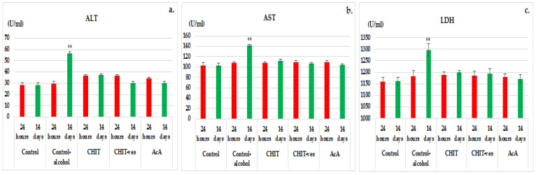
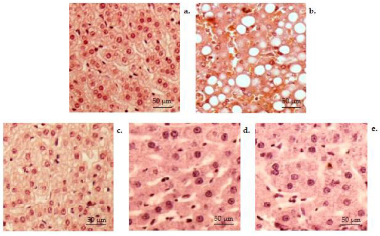
Background and Objectives In the past few decades, the studies concerning the natural polysaccharide chitosan have been centered on a new direction: its hepatoprotective action. The aim of our study was to evaluate the influence of previously designed chitosan lipid vesicles on the liver damage induced by alcohol consumption in mice. Materials and Methods The study involved the oral administration of substances in one daily dose as follows: Group 1 (control): water; Group 2 (control alcohol): 5% alcohol in water; Group 3 (CHIT): 0.1 mL/10 g body weight chitosan solution in animals treated with alcohol; Group 4 (CHIT-ves): 0.1 mL/10 g body chitosan vesicles in animals treated with alcohol; Group 5 (AcA): 200 mg/kg body ascorbic acid in animals treated with alcohol. In order to evaluate liver damage after alcohol consumption, the following hematological parameters were tested: the activity of alanine aminotransferase, aspartate aminotransferase and lactate dehydrogenase; serum values of urea and creatinine; the phagocytic capacity of polymorphonuclear neutrophilsin peripheral blood;serum opsonic capacity;bactericidal capacity of peritoneal macrophages; and the activity of malondialdehyde, glutathione peroxidase, superoxide dismutase and lactate dehydrogenase. Results and Conclusions The treatment with chitosan vesicles decreased liver enzyme activity and reduced the oxidative stress disturbances in alcoholic mice, thus repairing the hepatic functional and structural damages. These beneficial activities of chitosan vesicles were comparable with ascorbic acid effects in alcoholic mice.
Keywords: alcohol; chitosan; hepatoprotective; lipid vesicles; mice.
Conflict of interest statement
The authors declare no conflict of interest.
Figures


Similar articles
-
Reduction of the DNA damages, Hepatoprotective Effect and Antioxidant Potential of the Coconut Water, ascorbic and Caffeic Acids in Oxidative Stress Mediated by Ethanol.An Acad Bras Cienc. 2017 Apr-Jun;89(2):1095-1109. doi: 10.1590/0001-3765201720160581. Epub 2017 May 15. An Acad Bras Cienc. 2017. PMID: 28513780
-
Hepatoprotective effect of chitosan-caffeic acid conjugate against ethanol-treated mice.Exp Toxicol Pathol. 2017 Oct 2;69(8):618-624. doi: 10.1016/j.etp.2017.05.009. Epub 2017 Jun 7. Exp Toxicol Pathol. 2017. PMID: 28599809
-
Anti-Aging Effect of Chitosan Oligosaccharide on d-Galactose-Induced Subacute Aging in Mice.Mar Drugs. 2018 May 24;16(6):181. doi: 10.3390/md16060181. Mar Drugs. 2018. PMID: 29794973 Free PMC article.
-
Ganoderic acid A from Ganoderma lucidum protects against alcoholic liver injury through ameliorating the lipid metabolism and modulating the intestinal microbial composition.Food Funct. 2022 May 23;13(10):5820-5837. doi: 10.1039/d1fo03219d. Food Funct. 2022. PMID: 35543349
-
Hepatoprotective effects of Methyl ferulic acid on alcohol-induced liver oxidative injury in mice by inhibiting the NOX4/ROS-MAPK pathway.Biochem Biophys Res Commun. 2017 Nov 4;493(1):277-285. doi: 10.1016/j.bbrc.2017.09.030. Epub 2017 Sep 7. Biochem Biophys Res Commun. 2017. PMID: 28890346
Cited by
-
BDNF-loaded chitosan-based mimetic mussel polymer conduits for repair of peripheral nerve injury.Front Cell Dev Biol. 2024 Jul 1;12:1431558. doi: 10.3389/fcell.2024.1431558. eCollection 2024. Front Cell Dev Biol. 2024. PMID: 39011392 Free PMC article.
-
Effects of Dietary Supplementation with Chitosan on the Muscle Composition, Digestion, Lipid Metabolism, and Stress Resistance of Juvenile Tilapia (Oreochromis niloticus) Exposed to Cadmium-Induced Stress.Animals (Basel). 2024 Feb 6;14(4):541. doi: 10.3390/ani14040541. Animals (Basel). 2024. PMID: 38396509 Free PMC article.
-
Fast and Sustained Axonal Growth by BDNF Released from Chitosan Microspheres.Mar Drugs. 2023 Jan 27;21(2):91. doi: 10.3390/md21020091. Mar Drugs. 2023. PMID: 36827132 Free PMC article.
-
Protective Effects of Wine Polyphenols on Oxidative Stress and Hepatotoxicity Induced by Acrylamide in Rats.Antioxidants (Basel). 2022 Jul 10;11(7):1347. doi: 10.3390/antiox11071347. Antioxidants (Basel). 2022. PMID: 35883838 Free PMC article.
References
-
- Aravamudhan A., Ramos D.M., Nada A.A., Kumbar S.G. Natural and Synthetic Biomedical Polymers. Elsevier Inc.; Amsterdam, The Netherlands: 2014. Natural Polymers: Polysaccharides and Their Derivatives for Biomedical Applications. - DOI
-
- Dash M., Chiellini F., Ottenbrite R.M., Chiellini E. Chitosan—A versatile semi-synthetic polymer in biomedical applications. Prog. Polym. Sci. 2011;36:981–1014. doi: 10.1016/j.progpolymsci.2011.02.001. - DOI
-
- Adhikari H.S., Garai A., Khanal C., Adhikari R., Yadav P.N. Imidazole-2-carboxaldehyde Chitosan Thiosemicarbazones and Their Copper(II) Complexes: Synthesis, Characterization and Antitumorigenic Activity against Madin-Darby Canine Kidney Cell Line. Asian J. Chem. 2021;33:969–976. doi: 10.14233/ajchem.2021.23078. - DOI
-
- Islam S., Bhuiyan M.A.R., Islam M.N. Chitin and Chitosan: Structure, Properties and Applications in Biomedical Engineering. J. Polym. Environ. 2017;25:854–866. doi: 10.1007/s10924-016-0865-5. - DOI
MeSH terms
Substances
LinkOut - more resources
Full Text Sources
Miscellaneous
