

Tracheal Glomus Tumor: A Case Report with CT Imaging Features
- PMID: 35744054
- PMCID: PMC9229945
- DOI: 10.3390/medicina58060791
Tracheal Glomus Tumor: A Case Report with CT Imaging Features
Abstract
Background and objectives: Glomus tumors are rare benign tumors. The majority of them affect the skin the most and are rarer in the trachea, where the glomus bodies may not be present. Only scarce reports of tracheal glomus tumors have been reported solely with case reports of relevant articles.
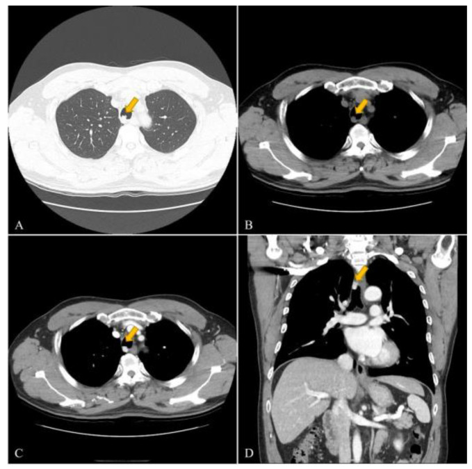
Materials and methods: A 53-year-old man, with a free previous medial history, presented to our hospital with tracheal mass which was incidentally found. He did not complain of any specific symptoms associated with the tracheal tumor. The contrast-enhanced chest computed tomography (CT) revealed an avid enhancing nodular lesion, which is similar to blood vessels, in the trachea, 3 cm above the carina level without definite airway obstruction.
Results: Successful tracheal resection and end-to-end anastomosis were performed on the patients; therefore, the final post-operative pathologic findings revealed a benign tracheal glomus tumor. The follow-up CT scan four months after operation showed complete removal of the tumor.
Conclusion: Tracheal glomus tumors, even rare entities, could be considered as a differential diagnosis if a highly enhancing mass appears on CT images.
Keywords: bronchoscopic biopsy; computed tomography.
Conflict of interest statement
The authors declare no conflict of interest.
Figures


Similar articles
-
Glomus tumor of the trachea.Asian Cardiovasc Thorac Ann. 2015 Mar;23(3):325-7. doi: 10.1177/0218492314528184. Epub 2014 Apr 2. Asian Cardiovasc Thorac Ann. 2015. PMID: 24696105
-
Successful resection of a glomus tumor arising from the lower trachea: report of a case.Surg Today. 1998;28(3):332-4. doi: 10.1007/s005950050134. Surg Today. 1998. PMID: 9548322
-
Glomus tumor of the trachea.Ann Thorac Surg. 2001 Aug;72(2):598-600. doi: 10.1016/s0003-4975(00)02278-5. Ann Thorac Surg. 2001. PMID: 11515904
-
Malignant glomus tumor of trachea: a case report with literature review.Asian Cardiovasc Thorac Ann. 2016 Jan;24(1):104-6. doi: 10.1177/0218492315608546. Epub 2015 Sep 28. Asian Cardiovasc Thorac Ann. 2016. PMID: 26420909 Review.
-
Successful resection of a glomus tumor of the trachea.Gen Thorac Cardiovasc Surg. 2011 Dec;59(12):815-8. doi: 10.1007/s11748-010-0772-y. Epub 2011 Dec 16. Gen Thorac Cardiovasc Surg. 2011. PMID: 22173681 Review.
Cited by
-
Extramural recurrence of tracheal glomus tumour following resection by rigid bronchoscopy.Respirol Case Rep. 2024 Feb 13;12(2):e01302. doi: 10.1002/rcr2.1302. eCollection 2024 Feb. Respirol Case Rep. 2024. PMID: 38351921 Free PMC article.
-
Diagnostic and Therapeutic Insights into Spinal Glomangioma of a Unique Intradural, Extramedullary Presentation-Systematic Review.Diseases. 2024 Jun 20;12(6):132. doi: 10.3390/diseases12060132. Diseases. 2024. PMID: 38920564 Free PMC article. Review.
-
Non-Surgical Treatment of Tracheal Glomus Tumour Using Rigid Fiberoptic Bronchoscopy: A Case Report.Respirol Case Rep. 2025 Jun 19;13(6):e70236. doi: 10.1002/rcr2.70236. eCollection 2025 Jun. Respirol Case Rep. 2025. PMID: 40546266 Free PMC article.
-
Tracheal airway obstruction induced by a large glomangioma: discussion of management and literature review.BMJ Case Rep. 2024 Sep 28;17(9):e261481. doi: 10.1136/bcr-2024-261481. BMJ Case Rep. 2024. PMID: 39343460 Free PMC article. Review.
References
-
- Specht K., Antonescu C.R. WHO Classification of Tumours of Soft Tissue and Bone. 5th ed. International Agency for Research on Cancer (IARC); Lyon, France: 2020. Glomus tumour; pp. 179–181.
Publication types
MeSH terms
LinkOut - more resources
Full Text Sources
