

Evaluation of Shear Bond Strength between Resin Composites and Conventional Glass Ionomer Cement in Class II Restorative Technique-An In Vitro Study
- PMID: 35744350
- PMCID: PMC9230775
- DOI: 10.3390/ma15124293
Evaluation of Shear Bond Strength between Resin Composites and Conventional Glass Ionomer Cement in Class II Restorative Technique-An In Vitro Study
Abstract
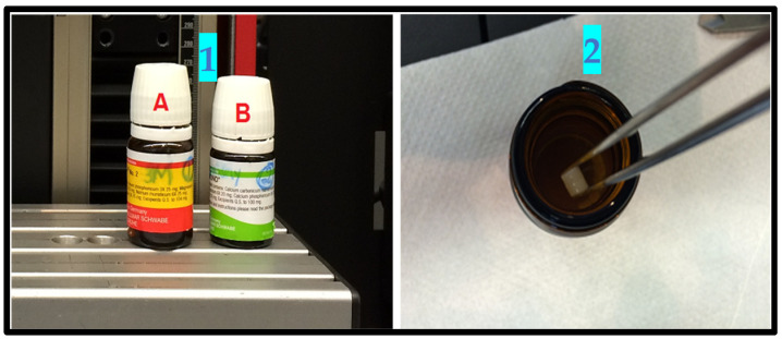
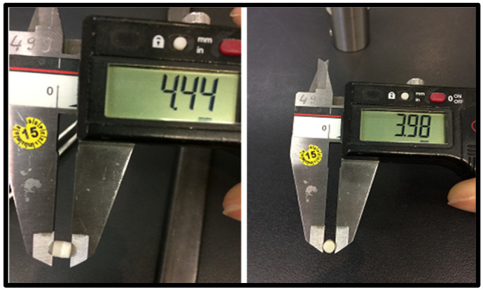
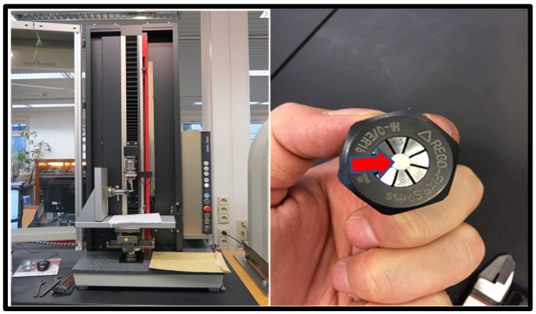
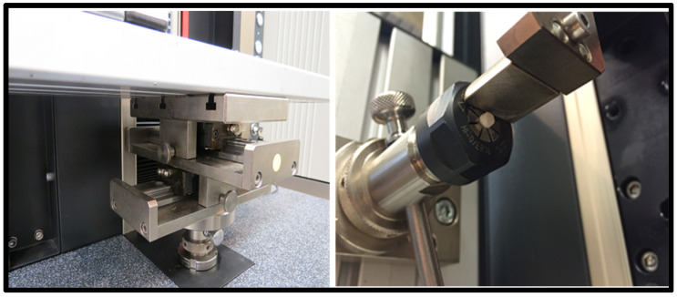
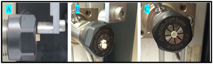
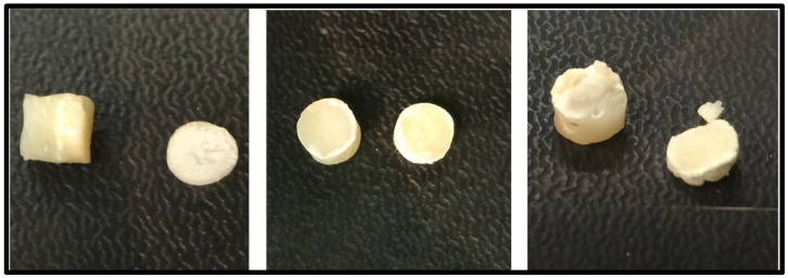
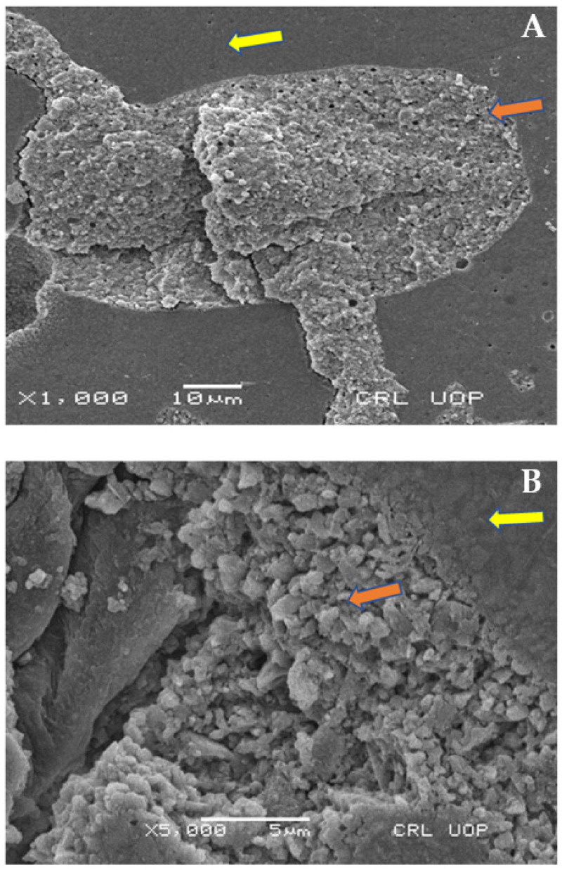
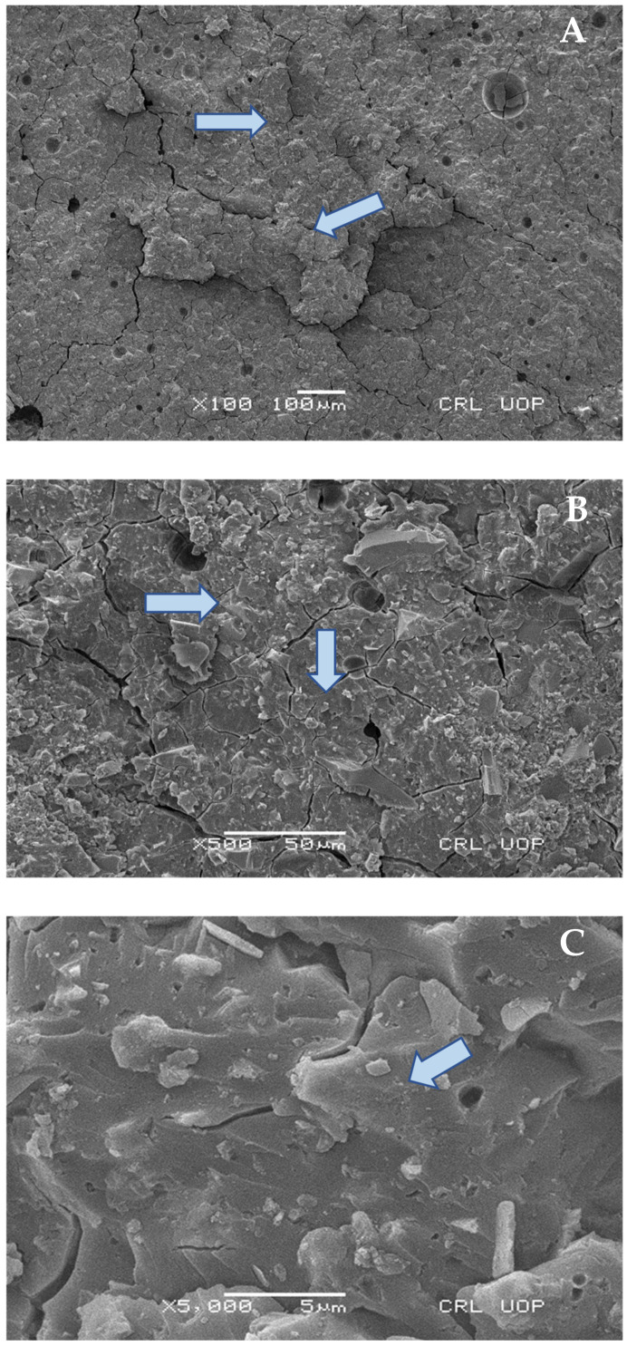
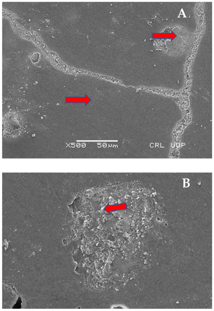
The success of dental restorations depends mainly on the ability to bond to other filling materials and tooth substances, in order to resist the multitude of forces acting on the bond within the oral cavity. Although the shortcomings of composite resins have been significantly reduced over the past three decades, microleakage due to shrinkage under masticatory loads is unavoidable. In order to overcome such problems, two materials laminated with matched properties can be used to achieve optimum results. The sandwich technique is an approach in which dentine is replaced by glass ionomer cement (GIC), and enamel is replaced by composite resin. In the past, numerous materials have been proposed with adequate properties to be used in this manner, but the results are conflicting in terms of bonding to the various forms of GIC, and the appearance of microcracks or gap formation during functional loading. This study aimed to evaluate the shear bond strength (SBS) and mode of failure between the following core materials: composite resins (CR) (Methacrylate Z350™, Ceram X™, and Spectrum™) with a base material of glass ionomer cement (GIC, Ketac Molar™). Eight samples were made with the help of polytetrafluoroethylene sheets (TEFLON, Wilmington, DE, USA). Each sheet consisted of holes which were 4 mm in diameter and 2 mm in thickness. The combination of materials was sandwiched. The samples were stored in distilled water and then placed in an incubator for 24 h in order to ensure complete polymerization. The samples were thermocycled for 500 cycles between 5-55 °C/ 30 s. Following thermocycling, SBS testing was performed using a universal testing machine. Additionally, scanning electron microscopy (SEM) was performed on representative samples for the bond failure analysis between GIC and the composite resins. The Ceram-X™ nanocomposite showed significantly higher bond strength than Methacrylate Z350™ or Spectrum™ (p = 0.002). The Methacrylate Z350™ and the Spectrum™ composite specimens demonstrated a similar SBS (p = 0.281). The SBS of the Ceram X™ to GIC was the highest compared to Methacrylate Z350™ and Spectrum™. Therefore Ceram X™ may produce a better bond with GIC, and may protect teeth against recurrent caries and failure of the restoration. Methacrylate Z350™ is comparable to Spectrum™ CR and can be used as an alternative. A combination of adhesive and mixed failure was observed in Methacrylate Z350™ CR and GIC, while adhesive failure was predominantly found in both Ceram X™ and Spectrum™ with GIC restorations.
Keywords: glass ionomer cement; microhybrid composites; nanocomposites; sandwich technique; shear bond strength.
Conflict of interest statement
All authors have declared that there are no conflict of interest.
Figures
References
-
- Latta M.A., Tsujimoto A., Takamizawa T., Barkmeier W.W. Enamel and dentin bond durability of self-adhesive restorative materials. J. Adhes. Dent. 2020;22:99–105. - PubMed
-
- Rathi S.D., Nikhade P., Chandak M., Motwani N., Rathi C., Chandak M. Microleakage in Composite Resin Restoration-A Review Article. J. Evol. Med. Dent. Sci. 2020;9:1006–1011. doi: 10.14260/jemds/2020/216. - DOI
Grants and funding
LinkOut - more resources
Full Text Sources
Miscellaneous
