. 2022 Jun 20;13(6):978.
doi: 10.3390/mi13060978.

Rational Design of Microfluidic Glaucoma Stent

Affiliations

Rational Design of Microfluidic Glaucoma Stent

Thomas Graf et al. Micromachines (Basel). .

Abstract

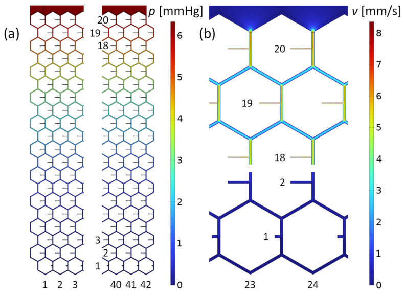
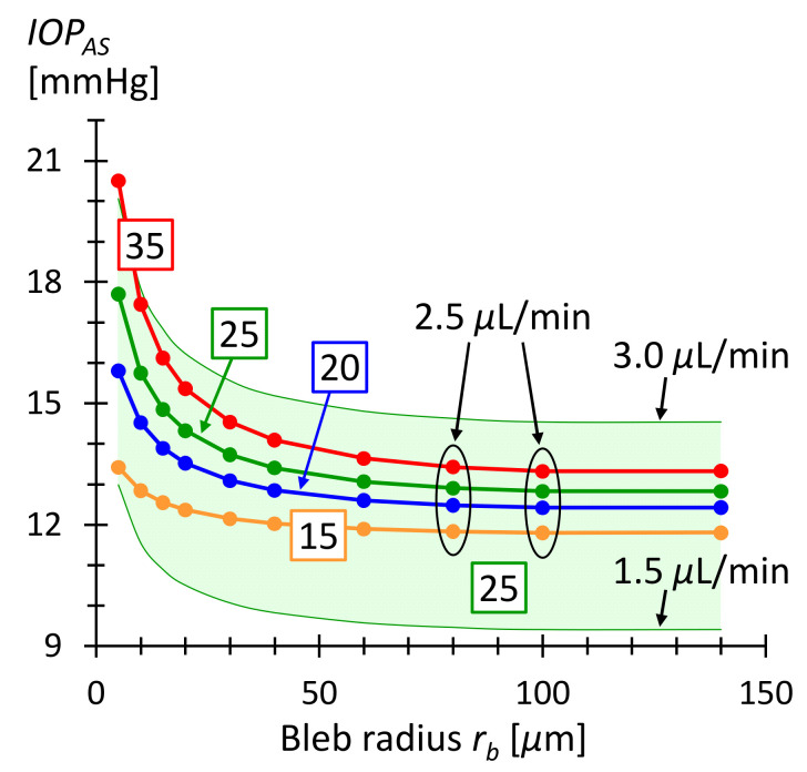
Glaucoma is a common, irreparable eye disease associated with high intraocular pressure. One treatment option is implantation of a stent to lower the intraocular pressure. A systematic approach to develop a microchannel stent meshwork that drains aqueous humor from the anterior chamber of the eye into the subconjunctival space is presented. The stent has a large number of outlets within its mesh structure that open into the subconjunctiva. The development approach includes a flow resistance model of the stent. Local adaption of the stent's tubular dimensions allows for adjustment of the flow resistance. In this way, an evenly distributed outflow into the subconjunctiva is achieved. We anticipate that microblebs will form at the stent outlets. Their size is crucial for drainage and control of intraocular pressure. An analytical model for bleb drainage is developed based on the porous properties of the subconjunctival tissue. Both models-the stent flow resistance model and the bleb drainage model-are verified by numerical simulation. The models and numerical simulation are used to predict intraocular pressure after surgery. They allow for a systematic and personalized design of microchannel stents. Stents designed in this way can stabilize the intraocular pressure between an upper and lower limit.

Keywords: IOP stabilization; bleb drainage; flow resistance model; glaucoma; microfluidic stent.


Conflict of interest statement

The authors declare no conflict of interest.

Figures

Figure A1
Figure A1
Various possible unit cells of the tubular hexagonal mesh are shown as shaded rectangles. The total resistance R0 in each unit cell is the same.
Figure 1
Figure 1
(a) Sectional anatomy of the eye with glaucoma stent. Part A is placed in the anterior chamber to collect AH. Part B conducts the AH into the meshwork of part C. Part C drains the fluid into the subconjunctival tissue. (b) Drainage pathways and flow balance in the eye after surgery. AH is produced in the ciliary body and drained by the trabecular meshwork (TM) via Schlemm’s Canal, the uveoscleral pathway (UP) and the stent. At the stent outlets, blebs form in the subconjunctiva on the scleral barrier. The indicated flow rates were used in numerical simulation.
Figure 2
Figure 2
The meshwork consists of honeycomb cells and is 12.6 mm wide and 5.2 mm high. Each hexagonal segment is a microtube (microchannel) with a square internal cross-section 14 μm wide. Each honeycomb cell contains an outlet tube with specific dimensions and a corresponding flow resistance (see enlarged detail). The black numbers are the dimensions; the red numbers denote the columns and rows of the meshwork. The liquid flows evenly from part B above into the meshwork.
Figure 3
Figure 3
Tubular structure of the stent and fluid resistance model. (a) The honeycomb meshwork geometry is the same everywhere; only the outlet tubes have different dimensions. The star-shaped part, colored in red, can be expressed as a fluidic resistance R0 between two subsequent outlet tubes. (b) Equivalent circuit diagram of a single column of the stent, from the fluid inlet (top) to the individual outlets. The numbered resistors R1 to R20 correspond to the various outlet tubes along the stent column. Due to the different dimensions, the outlet tubes have distinct flow resistances. The dimensions are chosen so that the same rate of liquid flows out of each tube. The diagram in (c) illustrates how the resistance R0 can be calculated using the flow resistance R of a straight channel segment (marked in blue). Drawing (d) depicts the relevant geometric entities for the calculation of the flow resistance.
Figure 4
Figure 4
Pressure and flow velocity field in the microchannel mesh computed with the program COMSOL. The boundary conditions were the inlet flow rate of 1.7μLmin and the outlet pressure of 0 mmHg at each orifice. (a) Pressure distribution in the stent of the leftmost and rightmost part of the mesh. The maximum value is 6.3 mmHg at the inlet, above row 20. (b) Flow velocities in the midplane of the meshwork. Shown are flow details at top and bottom of the stent, along the centerline of the honeycomb structure.
Figure 5
Figure 5
(a) COMSOL simulation of the outflow from a bleb with radius rb=30 μm. The inlet boundary condition is the mass flow rate M˙=Q˙b·ϱ=3.3×10¹¹kgs at the hemispherical bleb surface. This corresponds to Q˙b=Q˙t=2.0nLmin, given in Table 2. The outer boundary condition is the pressure pd=0.17 mm Hg at the domain boundary at rd=300 μm. pd is calculated by means of Equations (8) and (11) and used to mimic an infinite domain. The white lines and arrows indicate the flow direction given by the simulation. The axis of rotational symmetry is shown as a vertical, dashed red line. (b) Pressure as a function of distance from the origin. The blue line is the COMSOL result; the red circles are obtained using Equations (8) and (11).
Figure 6
Figure 6
(a) Microbleb array of 12.6×5.2 mm2 of the current stent meshwork. The microblebs lie on the scleral barrier and are spaced 300 µm apart. All blebs have a radius of rb=30 μm. AH flows from the bleb surfaces into the 0.6 mm thick subconjunctiva. (b,c) Drainage pressure field in the subconjunctival tissue determined by COMSOL simulation. The total flow rate of the bleb array is 1.7μLmin. Figure 6b shows a quarter of the simulation domain. Figure 6c is an enlarged view of 6b. The pressure inside the blebs is 7 mmHg and reaches 4.1 mmHg at the surface of the subconjunctiva. The pressure decreases rapidly in the plane of the array within the characteristic length 1/C=0.7 mm.
Figure 7
Figure 7
Drainage resistance Ra obtained by COMSOL simulations of hexagonal arrays as in Figure 6. (a) Ra as a function of bleb spacing da. The bleb radius is constant rb=30 μm. The curve reaches the value of 1.5 mmHgminμL for large separations, which is consistent with Equation (13) for free blebs. (b) Ra  as a function of bleb radius rb. The bleb spacing is constant at da=300 μm. For bleb radii rb>30 μm, the curve terminates in a constant value corresponding to the drainage resistance of a shallow macrobleb of 12.6×5.2 mm2 surface area.
Figure 8
Figure 8
IOPAS as a function of microbleb radius rb. The bleb separation is da=300 μm corresponding to the geometric period in the current honeycomb design. The numbers in the boxes are the IOP values before surgery in units of mmHg. The curves were calculated using Equation (16) and Q˙. IOPAS ranges from 12 and 15 mmHg for bleb radii greater than 30 μm. The green shaded area corresponds to the IOPAS for AH production rate varying between 1.5 and 3.0 μL/min and an IOP before surgery of 25 mmHg. The green shaded area demonstrates that the IOP is within a healthy range even with daily fluctuating production rates [4].

References

    1. Allison K., Patel D., Alabi O. Epidemiology of Glaucoma: The Past, Present, and Predictions for the Future. Cureus. 2020;12:e11686. doi: 10.7759/cureus.11686. - DOI - PMC - PubMed
    1. Zhang N., Wang J., Li Y., Jiang B. Prevalence of primary open angle glaucoma in the last 20 years: A meta-analysis and systematic review. Sci. Rep. 2021;11:13762. doi: 10.1038/s41598-021-92971-w. - DOI - PMC - PubMed
    1. Tham Y.C., Li X., Wong T.Y., Quigley H.A., Aung T., Cheng C.Y. Global prevalence of glaucoma and projections of glaucoma burden through 2040: A systematic review and meta-analysis. Ophthalmology. 2014;121:2081–2090. doi: 10.1016/j.ophtha.2014.05.013. - DOI - PubMed
    1. Lee R.M.H., Bouremel Y., Eames I., Brocchini S., Khaw P.T. Translating Minimally Invasive Glaucoma Surgery Devices. Clin. Transl. Sci. 2020;13:14–25. doi: 10.1111/cts.12660. - DOI - PMC - PubMed
    1. Do A.T., Parikh H., Panarelli J.F. Subconjunctival microinvasive glaucoma surgeries: An update on the Xen gel stent and the PreserFlo MicroShunt. Curr. Opin. Ophthalmol. 2020;31:132–138. doi: 10.1097/ICU.0000000000000643. - DOI - PubMed

LinkOut - more resources