Hyphal Fusions Enable Efficient Nutrient Distribution in Colletotrichum graminicola Conidiation and Symptom Development on Maize
- PMID: 35744664
- PMCID: PMC9231406
- DOI: 10.3390/microorganisms10061146
Hyphal Fusions Enable Efficient Nutrient Distribution in Colletotrichum graminicola Conidiation and Symptom Development on Maize
Abstract
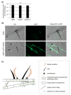
Hyphal and germling fusion is a common phenomenon in ascomycetous fungi. Due to the formed hyphal network, this process enables a coordinated development as well as an interaction with plant hosts and efficient nutrient distribution. Recently, our laboratory work demonstrated a positive correlation between germling fusion and the formation of penetrating hyphopodia on maize leaves outgoing from Colletotrichum graminicola oval conidia. To investigate the probable interconnectivity of these processes, we generated a deletion mutant in Cgso, in which homologs are essential for cellular fusion in other fungal species. However, hyphopodia development was not affected, indicating that both processes are not directly connected. Instead, we were able to link the cellular fusion defect in ∆Cgso to a decreased formation of asexual fruiting bodies of C. graminicola on the leaves. The monitoring of a fluorescent-labelled autophagy marker, eGFP-CgAtg8, revealed a high autophagy activity in the hyphae surrounding the acervuli. These results support the hypothesis that the efficient nutrient transport of degraded cellular material by hyphal fusions enables proper acervuli maturation and, therefore, symptom development on the leaves.
Keywords: Colletotrichum graminicola; autophagy; conidiation; falcate conidia; germling fusion; oval conidia.
Conflict of interest statement
The authors declare no conflict of interest. The funders had no role in the design of the study, the conducted analyses, the interpretation of data, the writing of the manuscript, or in the decision to publish the results.
Figures


Similar articles
-
Specialized infection strategies of falcate and oval conidia of Colletotrichum graminicola.Fungal Genet Biol. 2019 Dec;133:103276. doi: 10.1016/j.fgb.2019.103276. Epub 2019 Sep 21. Fungal Genet Biol. 2019. PMID: 31550526
-
The Colletotrichum graminicola striatin orthologue Str1 is necessary for anastomosis and is a virulence factor.Mol Plant Pathol. 2016 Aug;17(6):931-42. doi: 10.1111/mpp.12339. Epub 2016 Feb 18. Mol Plant Pathol. 2016. PMID: 26576029 Free PMC article.
-
Root infection and systemic colonization of maize by Colletotrichum graminicola.Appl Environ Microbiol. 2008 Feb;74(3):823-32. doi: 10.1128/AEM.01165-07. Epub 2007 Dec 7. Appl Environ Microbiol. 2008. PMID: 18065625 Free PMC article.
-
Gcc1 homologs regulate growth, oxidative stress, conidiation and appressorium formation in Colletotrichum siamense and Colletotrichum graminicola.Microb Pathog. 2023 Sep;182:106249. doi: 10.1016/j.micpath.2023.106249. Epub 2023 Jul 10. Microb Pathog. 2023. PMID: 37437644
-
The Small Ras Superfamily GTPase Rho4 of the Maize Anthracnose Fungus Colletotrichum graminicola Is Required for β-1,3-glucan Synthesis, Cell Wall Integrity, and Full Virulence.J Fungi (Basel). 2022 Sep 22;8(10):997. doi: 10.3390/jof8100997. J Fungi (Basel). 2022. PMID: 36294561 Free PMC article.
Cited by
-
Conserved perception of host and non-host signals via the a-pheromone receptor Ste3 in Colletotrichum graminicola.Front Fungal Biol. 2024 Oct 7;5:1454633. doi: 10.3389/ffunb.2024.1454633. eCollection 2024. Front Fungal Biol. 2024. PMID: 39435183 Free PMC article.
-
Decoding fungal communication networks: molecular signaling, genetic regulation, and ecological implications.Funct Integr Genomics. 2025 May 29;25(1):111. doi: 10.1007/s10142-025-01620-2. Funct Integr Genomics. 2025. PMID: 40439833 Review.
-
Long-distance gene flow and recombination shape the evolutionary history of a maize pathogen.IMA Fungus. 2025 Feb 21;16:e138888. doi: 10.3897/imafungus.16.138888. eCollection 2025. IMA Fungus. 2025. PMID: 40052074 Free PMC article.
References
-
- Bhunjun C.S., Phukhamsakda C., Jayawardena R.S., Jeewon R., Promputtha I., Hyde K.D. Investigating species boundaries in Colletotrichum. Fungal Divers. 2021;107:107–127. doi: 10.1007/s13225-021-00471-z. - DOI
-
- O’Connell R.J., Thon M.R., Hacquard S., Amyotte S.G., Kleemann J., Torres M.F., Damm U., Buiate E.A., Epstein L., Alkan N. Lifestyle transitions in plant pathogenic Colletotrichum fungi deciphered by genome and transcriptome analyses. Nat. Genet. 2012;44:1060–1065. doi: 10.1038/ng.2372. - DOI - PMC - PubMed
Grants and funding
LinkOut - more resources
Full Text Sources

