. 2022 Jun 6;10(6):1160.
doi: 10.3390/microorganisms10061160.

Structural and Functional Characterization of the Holliday Junction Resolvase RuvC from Deinococcus radiodurans

Affiliations

Structural and Functional Characterization of the Holliday Junction Resolvase RuvC from Deinococcus radiodurans

Chen Qin et al. Microorganisms. .

Abstract

Holliday junctions (HJs) are four-way DNA structures, which are an important intermediate in the process of homologous recombination. In most bacteria, HJs are cleaved by specific nucleases called RuvC resolvases at the end of homologous recombination. Deinococcus radiodurans is an extraordinary radiation-resistant bacterium and is known as an ideal model organism for elucidating DNA repair processes. Here, we described the biochemical properties and the crystal structure of RuvC from D. radiodurans (DrRuvC). DrRuvC exhibited an RNase H fold that belonged to the retroviral integrase family. Among many DNA substrates, DrRuvC specifically bound to HJ DNA and cleaved it. In particular, Mn2+ was the preferred bivalent metal co-factor for HJ cleavage, whereas high concentrations of Mg2+ inhibited the binding of DrRuvC to HJ. In addition, DrRuvC was crystallized and the crystals diffracted to 1.6 Å. The crystal structure of DrRuvC revealed essential amino acid sites for cleavage and binding activities, indicating that DrRuvC was a typical resolvase with a characteristic choice for metal co-factor.

Keywords: Deinococcus radiodurans; Holliday junction; Mn2+; RuvC.


Conflict of interest statement

The authors declare no conflict of interest.

Figures

Figure 1
Figure 1
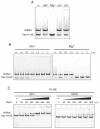
Protein purification and substrate specificities of DrRuvC. (A) Size exclusion chromatogram (Superdex 75 10/300 GL column) of recombinant DrRuvC protein. SDS-PAGE of representative eluted fractions is shown in inset. (B) Bandshift analysis of Holliday junctions (HJ-0X and HJ-12X). (C) Bandshift analysis of the indicated DNA structures. From left to right, the binding reactions contained 0, 50, 100, 200, 300, 400, 500 nM of DrRuvC protein. (D) Native PAGE analysis of DrRuvC cleavage of the indicated DNA structures.
Figure 2
Figure 2
Effect of metal ions on the resolvase activity of DrRuvC. (A) The Holliday junction cleavage assays were performed in the absence or presence of indicated metal ions. Substrate uses HJ-12X with 12 bp homologous core. 500 μM of each metal ion was used. (B) Metal ion titration experiment of DrRuvC cleavage activity.
Figure 3
Figure 3
Effect of metal ions on the HJ binding of DrRuvC. (A) The Holliday junction binding abilities of DrRuvC (250 nM) in the absence or presence of indicated metal ions (2 mM). The substrate HJ-0X with non-homologous core was used. (B) Mn2+ and Mg2+ titration experiments of HJ binding. (C) The binding abilities of DrRuvC to HJ-0X in the presence of 1 mM Mn2+ or in the absence of bivalent metal ions.
Figure 4
Figure 4
Crystal structure of DrRuvC. (A) Cartoon diagram of the overall structure of DrRuvC. The secondary structural elements are labeled. The N- and C-termini are indicated. (B) Structural superimposition of DrRuvC with EcRuvC and TtRuvC. (C) Electrostatic surface of the DrRuvC dimer colored according to the electrostatic surface potential (red: −1 kT/e to blue: +1 kT/e), shown in two orientations rotated by 180°. The location of the active sites is indicated by arrows.
Figure 5
Figure 5
The active sites of DrRuvC. (A) The active site of DrRuvC. DrRuvC is shown in grey and the catalytic residues are highlighted in magenta sticks. (B) Holliday junction cleavage assay of DrRuvC wild type (WT) or various mutants. (C) Superposition of the catalytic residues from DrRuvC (green) and EcRuvC (blue).

References

    1. Wright W.D., Shah S.S., Heyer W.-D. Homologous recombination and the repair of DNA double-strand breaks. J. Biol. Chem. 2018;293:10524–10535. doi: 10.1074/jbc.TM118.000372. - DOI - PMC - PubMed
    1. Mehta A., Haber J.E. Sources of DNA Double-Strand Breaks and Models of Recombinational DNA Repair. Cold Spring Harb. Perspect. Biol. 2014;6:a016428. doi: 10.1101/cshperspect.a016428. - DOI - PMC - PubMed
    1. Holliday R. A mechanism for gene conversion in fungi (Reprinted) Genet. Res. 2007;89:285–307. doi: 10.1017/S0016672308009476. - DOI - PubMed
    1. Bond C.S., Kvaratskhelia M., Richard D., White M.F., Hunter W.N. Structure of Hjc, a Holliday junction resolvase, from Sulfolobus solfataricus. Proc. Natl. Acad. Sci. USA. 2001;98:5509–5514. doi: 10.1073/pnas.091613398. - DOI - PMC - PubMed
    1. Hadden J.M., Declais A.-C., Carr S.B., Lilley D.M.J., Phillips S.E.V. The structural basis of Holliday junction resolution by T7 endonuclease. Nature. 2007;449:621-U15. doi: 10.1038/nature06158. - DOI - PubMed

LinkOut - more resources