. 2022 Jun 8;27(12):3684.
doi: 10.3390/molecules27123684.

Tripeptide IRW Protects MC3T3-E1 Cells against Ang II Stress in an AT2R Dependent Manner

Affiliations

Tripeptide IRW Protects MC3T3-E1 Cells against Ang II Stress in an AT2R Dependent Manner

Nan Shang et al. Molecules. .

Abstract

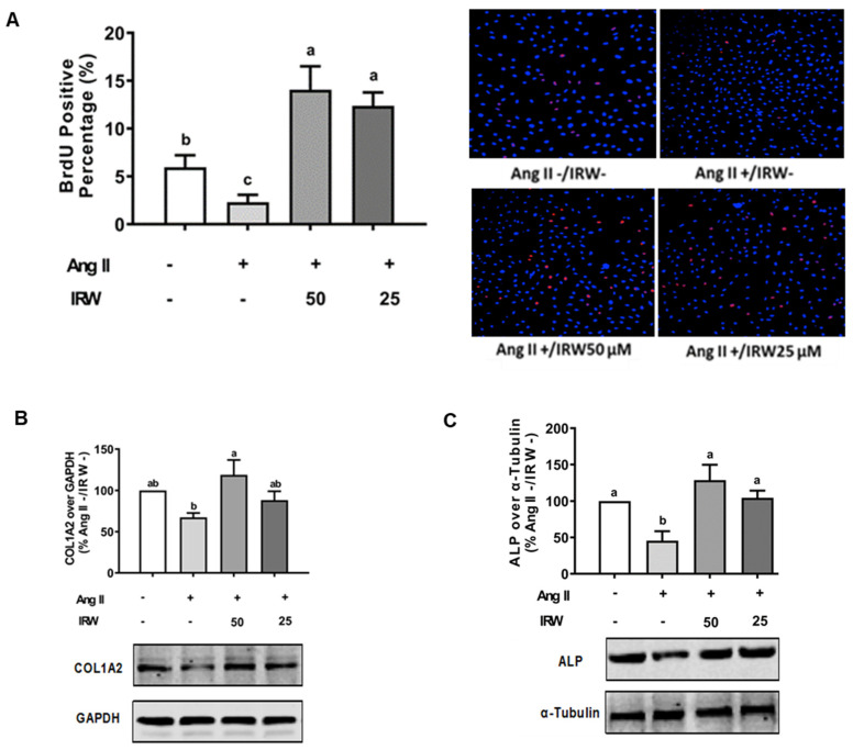
Multiple strategies including the use of bioactive peptides and other nutraceuticals are being adopted to maintain bone health. This study provides an improved and deeper understanding of the pharmacological effects that a bioactive peptide IRW (Ile-Arg-Trp) extends on bone health. Our results showed that IRW treatment protects osteoblasts against Ang II induced decline in cell proliferation and restores protein levels of collagen type I alpha 2 chain (COL1A2) and alkaline phosphatase (ALP) levels in MC3T3-E1 cells (p < 0.05). Apart from augmentation of these mineralization factors, the angiotensin II (Ang II) induced apoptotic stress in osteoblasts was mitigated by IRW as well. At the molecular level, IRW abolished the cytochrome-c release via modulation of pro-and anti-apoptotic genes in MC3T3-E1 cells (p < 0.05). Interestingly, IRW also increased cellular levels of cytoprotective local RAAS factors such as MasR, Ang (1−7), ACE2, and AT2R, and lowered the levels of Ang II effector receptor (AT1R). Further, our results indicated a lower content of inflammation and osteoclastogenesis biomarkers such as cyclooxygenase 2 (COX2), nuclear factor kappa B (NF-κB), and receptor activator of nuclear factor kappa-B ligand (RANKL) following IRW treatment in MC3T3-E1 cells (p < 0.05). The use of an antagonist-guided cell study indicated that IRW contributed to the process of cytoprotection and proliferation of osteoblasts via Runt-related transcription factor 2 (RUNX2) in face of Ang II stress in an AT2R dependent manner. The key findings of our study showed that IRW could potentially have a therapeutic role in the treatment and/or prevention of bone disorders.

Keywords: AT2R; IRW; RAAS; RUNX2; osteoporosis; peptides.


Conflict of interest statement

The authors declare no conflict of interest.

Figures

Figure 1
Figure 1
The effect of IRW on promoting osteoblastic activity against Ang II stimulation. (A) Osteoblast cells MC3T3-E1 were pre-treated with IRW (50 μM and 25 μM) for 2 h prior to Ang II (1 μM) and then co-cultured for 24 h. BrDU incorporation assay (A) was performed as described in methodology and the BrDU positive cells were counted; (BH) osteoblast cells MC3T3-E1 were pre-treated with IRW (50 μM and 25 μM) for 2 h prior to Ang II (1 μM) and then co-cultured for 24 h. Whole cell lysates were used for the Western blotting analysis of COL1A2 (B), ALP (C), RANKL (D), and OPG (E). (F) Osteoblast cells MC3T3-E1 were cultured in mineralization medium (MEM-α medium with ascorbic acid and β-glycerophosphate) and treated with IRW (50 μM and 25 μM) and Ang II (1 μM) for 15 days. Alizarin Red staining was performed, and the images were captured. (G,H) Osteoblast cells MC3T3-E1 were pre-treated with IRW (50 μM and 25 μM) for 2 h prior to Ang II (1 μM) and then co-cultured for 24 h. Both the culture medium (G) and whole cell lysates (H) were collected for ALP activity analysis. All results are representative of 4~6 independent experiments and expressed as mean ± SEM. Mean without a common letter indicated p < 0.05.
Figure 1
Figure 1
The effect of IRW on promoting osteoblastic activity against Ang II stimulation. (A) Osteoblast cells MC3T3-E1 were pre-treated with IRW (50 μM and 25 μM) for 2 h prior to Ang II (1 μM) and then co-cultured for 24 h. BrDU incorporation assay (A) was performed as described in methodology and the BrDU positive cells were counted; (BH) osteoblast cells MC3T3-E1 were pre-treated with IRW (50 μM and 25 μM) for 2 h prior to Ang II (1 μM) and then co-cultured for 24 h. Whole cell lysates were used for the Western blotting analysis of COL1A2 (B), ALP (C), RANKL (D), and OPG (E). (F) Osteoblast cells MC3T3-E1 were cultured in mineralization medium (MEM-α medium with ascorbic acid and β-glycerophosphate) and treated with IRW (50 μM and 25 μM) and Ang II (1 μM) for 15 days. Alizarin Red staining was performed, and the images were captured. (G,H) Osteoblast cells MC3T3-E1 were pre-treated with IRW (50 μM and 25 μM) for 2 h prior to Ang II (1 μM) and then co-cultured for 24 h. Both the culture medium (G) and whole cell lysates (H) were collected for ALP activity analysis. All results are representative of 4~6 independent experiments and expressed as mean ± SEM. Mean without a common letter indicated p < 0.05.
Figure 2
Figure 2
The effect of IRW on preventing osteoblast cell apoptosis against Ang II stimulation. Osteoblast cells MC3T3-E1 were pre-treated with IRW (50 μM and 25 μM) for 2 h prior to Ang II (1 μM) and then co-cultured for 24 h. (A) Cells were collected, fixed, and the cell apoptosis rate was measured by flow cytometry; (B) DHE staining was performed to measure the levels of oxidative stress; (CF) total RNA was extracted using TRIzol, converted to cDNA, and then the gene expression of mitochondrial apoptotic markers (C) Bcl-2, (D) Bax, (E) Caspase 3, and (F) Cytochrome C was measured by qPCR (quantitative PCR). All results are representative of 4~6 independent experiments and expressed as mean ± SEM. Mean without a common letter indicated p < 0.05.
Figure 3
Figure 3
The effect of IRW on regulating RAAS signaling with Ang II stimulation. Osteoblast cells MC3T3-E1 were pre-treated with IRW (50 μM and 25 μM) for 2 h prior to Ang II (1 μM) and then co-cultured for 24 h. Whole cell lysates were collected and used for the ELISA and Western blotting analysis. (A) The RIPA buffer extracts were used for measuring Ang (1–7) levels using ELISA and the protein expression of (B) angiotensin-converting enzyme 2 (ACE II), (C) angiotensin II receptor type 1 (AT1R), (D) angiotensin II receptor type 2 (AT2R), and (E) Mas receptor (MasR) were measured using Western blot. All results are representative of 6 independent experiments and expressed as mean ± SEM. Mean without a common letter indicated p < 0.05.
Figure 4
Figure 4
The effect of IRW on Cox2 and NF-κB p65 with Ang II stimulation. Osteoblast cells MC3T3-E1 were pre-treated with IRW (50 μM and 25 μM) for 2 h prior to Ang II (1 μM) and then co-cultured for 24 h. Whole cell lysates were collected and used for the Western blotting analysis to measure the protein expression of (A) cyclooxygenase-2 (Cox2) and (B) NF-κB p65. All results are representative of 6 independent experiments and expressed as mean ± SEM. Mean without a common letter indicated p < 0.05.
Figure 5
Figure 5
The impact of AT2R and MasR on cytoprotective activity of IRW against Ang II stimulation. Osteoblast cells MC3T3-E1 were pre-treated with IRW (50 μM and 25 μM) for 2 h prior to Ang II (1 μM) and then co-cultured for 24 h with or without AT2R (PD123319) and MasR (A779) inhibitors (A,C). Whole cell lysates were collected and used for the Western blotting analysis was conducted to measure the protein expression of (B,D) RUNX2. All results are representative of 3–6 independent experiments and expressed as mean ± SEM. Mean without a common letter indicated p < 0.05 while ** indicates p < 0.01.
Figure 6
Figure 6
Summary of cytoprotective and osteogenic impact of IRW against Ang II stimulation.

Similar articles

References

    1. Morgan E.F., Gerstenfeld L.C. Marcus and Feldman’s Osteoporosis. Elsevier; Amsterdam, The Netherlands: 2021. The bone organ system: Form and function; pp. 15–35.
    1. Bislev L.S., Sikjær T., Rolighed L., Rejnmark L. Relationship Between Aldosterone and Parathyroid Hormone, and the Effect of Angiotensin and Aldosterone Inhibition on Bone Health. Clin. Rev. Bone Miner. Metab. 2015;13:194–205. doi: 10.1007/s12018-015-9182-0. - DOI
    1. Högberg U., Andersson J., Högberg G., Thiblin I. Metabolic bone disease risk factors strongly contributing to long bone and rib fractures during early infancy: A population register study. PLoS ONE. 2018;13:e0208033. doi: 10.1371/journal.pone.0208033. - DOI - PMC - PubMed
    1. Mo C., Ke J., Zhao D., Zhang B. Role of the renin–angiotensin–aldosterone system in bone metabolism. J. Bone Miner. Metab. 2020;38:772–779. doi: 10.1007/s00774-020-01132-y. - DOI - PubMed
    1. Fountain J.H., Lappin S.L. Physiology, Renin Angiotensin System. StatPearls Publishing; Treasure Island, FL, USA: 2017. - PubMed

MeSH terms