Review
. 2022 Jun 18;27(12):3914.
doi: 10.3390/molecules27123914.

Near-Infrared-Emissive AIE Bioconjugates: Recent Advances and Perspectives

Affiliations
Review

Near-Infrared-Emissive AIE Bioconjugates: Recent Advances and Perspectives

Wenshuai Luo et al. Molecules. .

Abstract

Near-infrared (NIR) fluorescence materials have exhibited formidable power in the field of biomedicine, benefiting from their merits of low autofluorescence background, reduced photon scattering, and deeper penetration depth. Fluorophores possessing planar conformation may confront the shortcomings of aggregation-caused quenching effects at the aggregate level. Fortunately, the concept of aggregation-induced emission (AIE) thoroughly reverses this dilemma. AIE bioconjugates referring to the combination of luminogens showing an AIE nature with biomolecules possessing specific functionalities are generated via the covalent conjugation between AIEgens and functional biological species, covering carbohydrates, peptides, proteins, DNA, and so on. This perfect integration breeds unique superiorities containing high brightness, good water solubility, versatile functionalities, and prominent biosafety. In this review, we summarize the recent progresses of NIR-emissive AIE bioconjugates focusing on their design principles and biomedical applications. Furthermore, a brief prospect of the challenges and opportunities of AIE bioconjugates for a wide range of biomedical applications is presented.

Keywords: NIR emission; aggregation-induced emission; bioconjugates; biomedical applications.


Conflict of interest statement

The authors declare no conflict of interest.

Figures
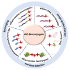
Figure 1
Figure 1
Schematic diagram of the representative bioconjugate coupling with NIR-emissive AIEgens.
Figure 2
Figure 2
(a) Structures of β-gal-targeting probes. (b) Schematic diagram of QM-HBT-β-gal towards β-gal. (c) Long-term tracking capability of QM-HBT-β-gal. Copyright 2019, Frontier Media.
Figure 3
Figure 3
(a) Chemical structures of enzyme-activatable AIEgen-peptide bioconjugates. (b) CTSE-responsive bioconjugate (QM-HSP-CPP) for PC cancer detection. (c) Schematic diagram of fluorescence tracking endogenous CTSE after intra-tumoral or intravenous injection of QM-HSP-CPP. Copyright 2022, Wiley-VCH.
Figure 4
Figure 4
(a) Chemical structure of targetable AIEgen-peptide bioconjugates. (b) Schematic illustration of the working mechanism of DBT-2FFGYSA. (c) CLSM images of PC-3 cancer cells incubated with DBT-2FFGYSA. Copyright 2021, Wiley-VCH. (d) Schematic diagram of the working mechanism of Q1-PEP. (e) Particle size of Q1-PEP NPs in deionized water. (f) Photostability of Q1-PEP nanodots compared to ER tracker. (g) Photostability of Q1-PEP nanodots in contrast to Cy 5.5 in vivo. Copyright 2018, Wiley-VCH.
Figure 5
Figure 5
(a) Chemical structures of AIEgens reported recently to conjugate with proteins. (b) Absorption and emission spectra of mAb-CSPP bioconjugate. (c) Absorption and emission spectra of mAb-Cy3 bioconjugate. (d) Bright-field and fluorescent images of HCC827 cancer cells under different treatments. Copyright 2017, Royal Society of Chemistry. (e) Structures of AIE810NP-chicken IgY/AIE810NP-SARS-CoV-2 antigen and the diagram of lateral flow immunoassay for the detection of IgM and IgG. (f) Interpretation of different test results. Notably, the invalid test strip herein demonstrated only represents one case of a test with the result ‘invalid’. (g) The value of IM/IC for the detection of IgM from 142 pre-COVID samples. (h) The value of IG/IC for the detection of IgG from 142 pre-COVID samples. (i) Sensitivity of IgM/IgG testing for 172 serum samples from COVID-infected patients. Copyright 2021, American Chemical Society.
Figure 6
Figure 6
(a) Synthetic route of TPE-R-DNA, TPE-R-AT, and the diagram of detecting MnSOD mRNA. (b) Size distributions of TPE-R-DNA (left) and TPE-R-AT (right). (c) Photoluminescence spectra of TPE-R-DNA in the presence of Exo III and different concentrations of MnSOD mRNA (0−1000 pM). (d) CLSM images of many cancer tissues and their HE staining images. (e) Fluorescent signal remaining percent of TPE-R-DNA in liver cancer tissue with increasing scanning numbers. (f) Fluorescence intensity of cancer tissues and their adjacent tissues. (g) CLSM images of renal cancer tissues (Ca) and their adjacent tissues (AT). Copyright, 2018, American Chemical Society.
Figure 7
Figure 7
(a) Chemical structures of vectors 1–4. (b) In vivo imaging of tumor-bearing mice at 24 h post-injection of vector 4/DNA NPs. (c) Ex vivo biodistribution of various organs and tumor tissue from tumor-bearing mice at 24 h post-injection with vector 4/DNA NPs. (d) HeLa cells stained by calcein-AM (green, live) and PI (red, dead). Copyright, 2021, American Chemical Society.
Figure 8
Figure 8
(a) Chemical structures of AIE-Pyo series and TVP-S. (b) Normalized fluorescence spectra of AIEgen-silks. (c) Fluorescence retention proportion of dyes after washing with soapy water. (d) Preparation and photos of WLE silk. (e) Two-photon fluorescent images of MTPABP-silk through the chicken tissues of 460 μm. Copyright 2021, Wiley-VCH. (f) Diagram of bacterial imaging, targeting, and killing driven by TVP-PAP. (g) Absorption spectra of TVP-PAP, PAP, and TVP-S. (h) DCFH for ROS detection of TVP-PAP. (i) Fluorescence imaging of P. aeruginosa and A. baumanni co-incubated with TVP-PAP for 30 min. (j) The survival rate and colonies of P. aeruginosa and A. baumanni co-incubated with TVP-PAP in the present light irradiation for 30 min. (k) The survival rates and colonies of P. aeruginosa and S. aureus co-incubated with TVP-PAP in the present light irradiation for 30 min. (l) The wound-healing rates at days 2, 5, and 8 under different treatments. Copyright, 2020, American Chemical Society.

Similar articles

Cited by

References

    1. Giepmans B.N.G., Adams S.R., Ellisman M.H., Tsien R.Y. The Fluorescent Toolbox for Assessing Protein Location and Function. Science. 2006;312:217–224. doi: 10.1126/science.1124618. - DOI - PubMed
    1. Lee M.H., Kim J.S., Sessler J.L. Small molecule-based ratiometric fluorescence probes for cations, anions, and biomolecules. Chem. Soc. Rev. 2015;44:4185–4191. doi: 10.1039/C4CS00280F. - DOI - PMC - PubMed
    1. Etzioni R., Urban N., Ramsey S., McIntosh M., Schwartz S., Reid B., Radich J., Anderson G., Hartwell L. The case for early detection. Nat. Rev. Cancer. 2003;3:243–252. doi: 10.1038/nrc1041. - DOI - PubMed
    1. Beatriz S.P., Luis N., Leonor C., Laura M., Elena M., Yolanda F.N. Imaging Techniques and Scanning Electron Microscopy as Tools for Characterizing a Si-Based Material Used in Air Monitoring Applications. Materials. 2016;9:109. doi: 10.3390/ma9020109. - DOI - PMC - PubMed
    1. Garg P.K., Singh S.K., Prakash G., Jakhetiya A., Pandey D. Role of positron emission tomography-computed tomography in non-small cell lung cancer. World J. Methodol. 2016;6:105–111. doi: 10.5662/wjm.v6.i1.105. - DOI - PMC - PubMed

Substances

LinkOut - more resources