. 2022 Jun 10;14(12):2411.
doi: 10.3390/nu14122411.

Alisol B Alleviates Hepatocyte Lipid Accumulation and Lipotoxicity via Regulating RARα-PPARγ-CD36 Cascade and Attenuates Non-Alcoholic Steatohepatitis in Mice

Affiliations

Alisol B Alleviates Hepatocyte Lipid Accumulation and Lipotoxicity via Regulating RARα-PPARγ-CD36 Cascade and Attenuates Non-Alcoholic Steatohepatitis in Mice

Zhuohui Zhao et al. Nutrients. .

Abstract

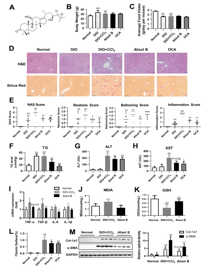
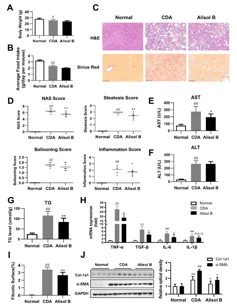
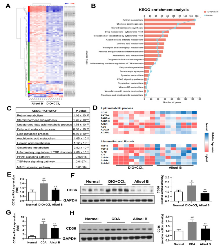
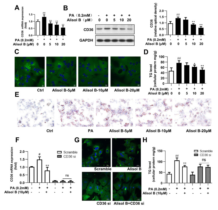
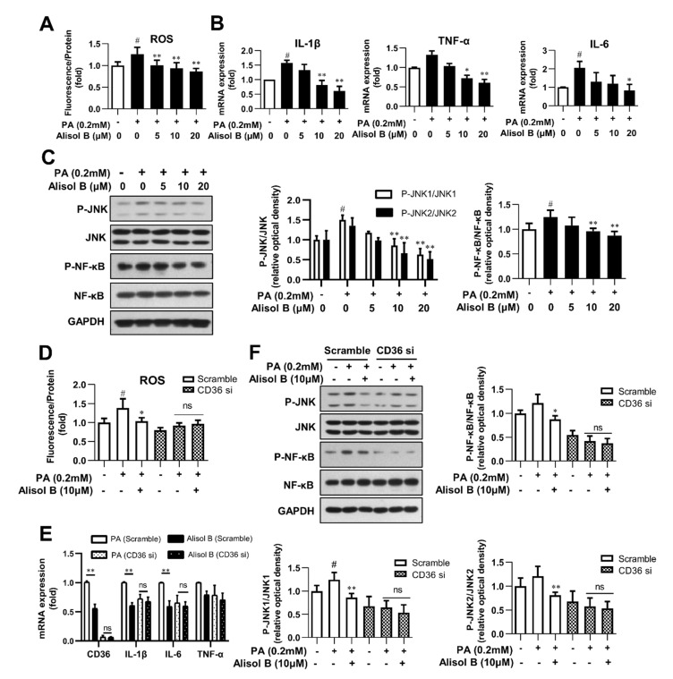
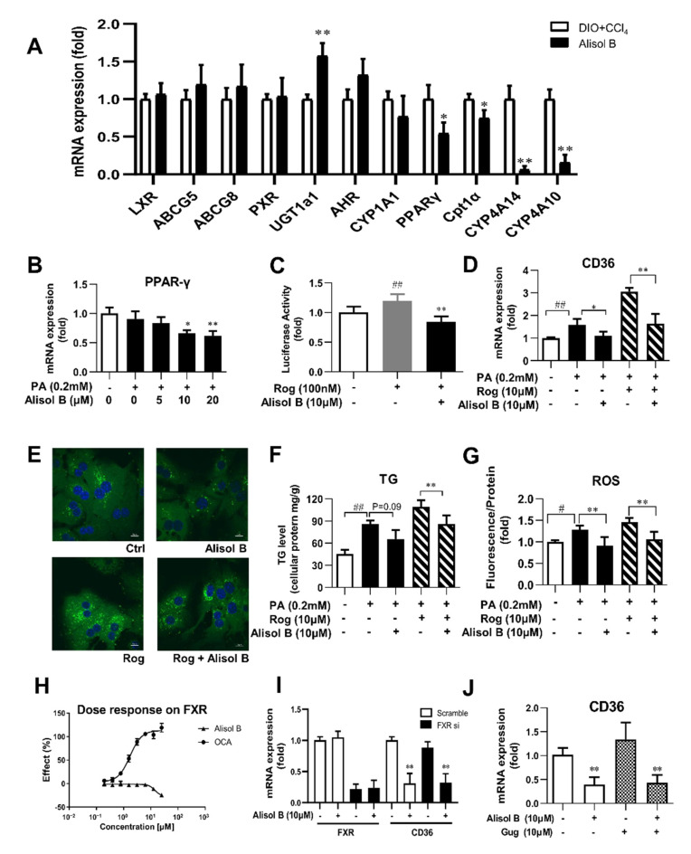
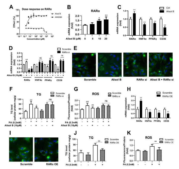
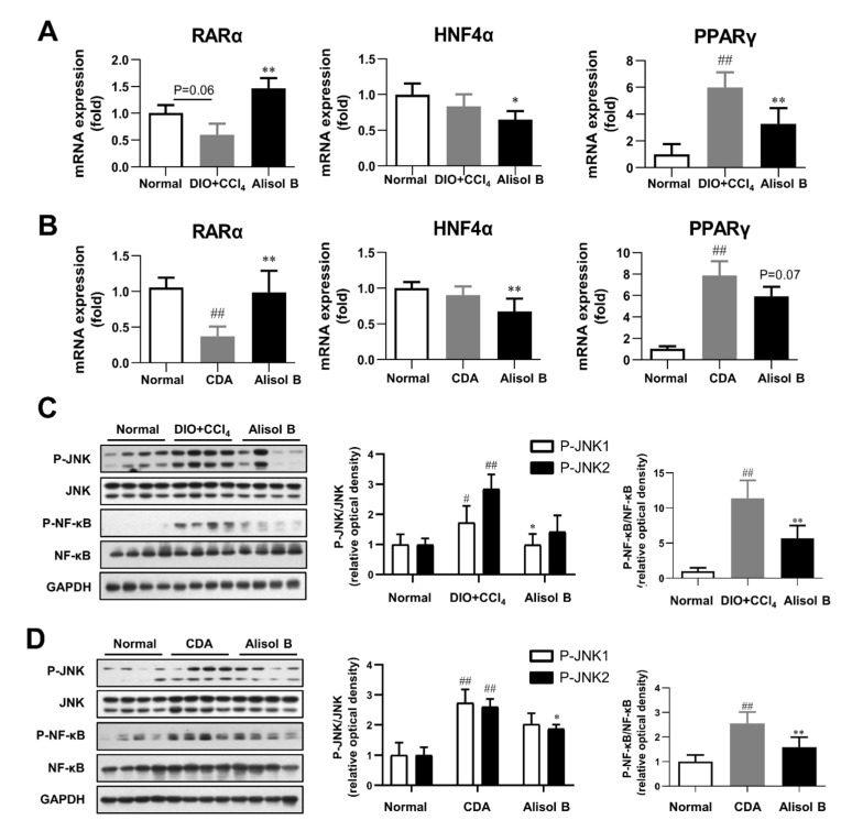
Non-alcoholic steatohepatitis (NASH) is a common chronic liver disease worldwide, with no effective therapies available. Discovering lead compounds from herb medicine might be a valuable strategy for the treatment of NASH. Here, we discovered Alisol B, a natural compound isolated from Alisma orientalis (Sam.), that attenuated hepatic steatosis, inflammation, and fibrosis in high-fat diet plus carbon tetrachloride (DIO+CCl4)-induced and choline-deficient and amino acid-defined (CDA)-diet-induced NASH mice. RNA-seq showed Alisol B significantly suppressed CD36 expression and regulated retinol metabolism in NASH mice. In mouse primary hepatocytes, Alisol B decreased palmitate-induced lipid accumulation and lipotoxicity, which were dependent on CD36 suppression. Further study revealed that Alisol B enhanced the gene expression of RARα with no direct RARα agonistic activity. The upregulation of RARα by Alisol B reduced HNF4α and PPARγ expression and further decreased CD36 expression. This effect was fully abrogated after RARα knockdown, suggesting Alisol B suppressed CD36 via regulating RARα-HNF4α-PPARγ cascade. Moreover, the hepatic gene expression of RARα was obviously decreased in murine NASH models, whereas Alisol B significantly increased RARα expression and decreased CD36 expression, along with the downregulation of HNF4α and PPARγ. Therefore, this study showed the unrecognized therapeutic effects of Alisol B against NASH with a novel mechanism by regulating RARα-PPARγ-CD36 cascade and highlighted Alisol B as a promising lead compound for the treatment of NASH.

Keywords: Alisol B; CD36; RARα; non-alcoholic steatohepatitis.


Conflict of interest statement

The authors declare no conflict of interest.

Figures

Figure 1
Figure 1
Alisol B ameliorated NASH in a DIO+CCl4-induced murine model. (A) The chemical structure of Alisol B. The therapeutic effects of Alisol B (100 mg/kg, once daily, p.o.) were evaluated in DIO+CCl4-induced mice as described in the Materials and Methods. (B) Body weight and (C) average food intake were measured. (D) H&E staining (200× magnification) and Sirius Red staining (100× magnification) were performed. (E) NAS score, steatosis score, ballooning score, and inflammation score were quantified. (F) Liver TG, (G) serum ALT and (H) AST, (I) hepatic mRNA levels of inflammatory cytokines, (J) hepatic MDA, and (K) GSH levels were measured. (L) Percentage of collagen deposition was calculated. (M) Western blot analysis of hepatic α-SMA and Col-1a1 expression was conducted and quantified. Data are presented as the mean ± SD, n = 8. # p < 0.05, ## p < 0.01 compared with the normal control group; * p < 0.05, ** p < 0.01 compared with the DIO+CCl4 group.
Figure 2
Figure 2
Alisol B ameliorated NASH in a CDA diet-induced murine model. The therapeutic effects of Alisol B (100 mg/kg, once daily, p.o.) were evaluated in CDA diet-fed mice as described in the Materials and Methods. (A) Body weight and (B) average food intake were measured. (C) H&E staining (200× magnification) and Sirius Red staining (100× magnification) were performed. (D) NAS score, steatosis score, ballooning score, and inflammation score were quantified. (E) Serum AST and (F) ALT levels, (G) liver TG, and (H) hepatic mRNA levels of inflammatory cytokines were measured. (I) Percentage of collagen deposition was calculated. (J) Western blot analysis of hepatic α-SMA and Col-1a1 was conducted and quantified. Data are expressed as mean ± SD, n = 8. # p < 0.05, ## p < 0.01 compared with the normal control group; * p < 0.05, ** p < 0.01 compared with the CDA group.
Figure 3
Figure 3
RNA-seq analysis. (A) Heat map, (B) KEGG enrichment analysis, (C) enriched KEGG biological pathway, and (D) kay genes expression involved in lipid metabolic process, inflammation, and fibrosis were performed in the liver of DIO+CCl4-induced mice. (E) RT-PCR analysis and (F) Western blot analysis of CD36 expression were conducted in DIO+CCl4-induced mice. (G) RT-PCR analysis and (H) Western blot analysis of CD36 expression were conducted in CDA-diet-fed mice. Data are expressed as mean ± SD, n = 8. # p < 0.05, ## p < 0.01 compared with the normal control group; ** p < 0.01 compared with the DIO+CCl4 group or CDA group.
Figure 4
Figure 4
Alisol B ameliorated cellular TG accumulation in primary hepatocytes by inhibiting FFA uptake in a CD36-dependent manner. Mouse primary hepatocytes were incubated with indicated concentration of Alisol B under PA (0.2 mM)-stimulated condition. (A) RT-PCR analysis and (B) Western blot analysis of CD36 expression, (C) BODIPY-C16 fluorescence (600× magnification), (D) cellular TG, and (E) Oil Red O staining (200× magnification) were conducted. (FH) Primary hepatocytes were pretreated with CD36 siRNA for 24 h and then treated with Alisol B (10 μM) under PA-induced conditions. (F) CD36 mRNA level, (G) BODIPY-C16 fluorescence (600× magnification), and (H) cellular TG were examined. Data are expressed as mean ± SD, n = 5. # p < 0.05, ## p < 0.01 compared with the normal control group; * p < 0.05, ** p < 0.01 compared with the PA-induced group.
Figure 5
Figure 5
Alisol B inhibited oxidative stress and inflammation in primary hepatocytes in a CD36-dependent manner. Mouse primary hepatocytes were incubated with indicated concentration of Alisol B under PA (0.2 mM)-stimulated conditions. (A) Cellular ROS level; (B) the mRNA levels of IL-1β, IL-6, and TNF-α; and (C) JNK1/2 and NF-κB phosphorylation were detected. (DF) Primary hepatocytes were pretreated with CD36 siRNA for 24 h and then treated with Alisol B (10 μM) under PA-induced conditions. (D) Cellular ROS; (E) the mRNA levels of IL-1β, IL-6, and TNF-α; and (F) JNK1/2 and NF-κB phosphorylation were detected. Data are expressed as mean ± SD, n = 5 for all of the groups, except for n = 6 in Figure (F). # p < 0.05 compared with the normal control group; * p < 0.05, ** p < 0.01 compared with the PA-induced group.
Figure 6
Figure 6
Alisol B suppressed CD36 expression through downregulating PPARγ, and this effect was independent of FXR. (A) The mRNA expression of hepatic LXR, PXR, AHR, PPARγ, and their target genes were examined in DIO+CCl4-induced NASH mice. (B) The gene expression of PPARγ was measured in PA-induced primary hepatocytes treated with Alisol B. (C) Huh7 cells were transiently transfected and then treated with Alisol B (10 μM) in the presence of Rosiglitazone. The transactivation activity of PPARγ was detected in luciferase reporter assay. (DG) Mouse primary hepatocytes were treated with Alisol B (10 μM) and Rosiglitazone (10 μM) under PA-stimulated conditions. (D) CD36 mRNA level, € BODIPY-C16 fluorescence (600× magnification), (F) cellular TG, and (G) cellular ROS were evaluated. (H) Huh7 cells were transiently transfected and then treated with Alisol B. The agonistic activity on FXR was detected in luciferase reporter assay. (I) The mRNA levels of FXR and CD36 in FXR knockdown hepatocytes treated with Alisol B (10 μM) were detected. (J) The mRNA level of CD36 in hepatocytes treated with Alisol B (10 μM) in the presence of Gugglusterone (10 μM) was detected. Data in (A) are expressed as mean ± SD, n = 8. * p < 0.05, ** p < 0.01 compared with DIO+CCl4 group. Data in (BH) are expressed as mean ± SD, n = 5. # p < 0.05, ## p < 0.01 compared with normal control group; * p < 0.05, ** p < 0.01 compared with the PA-induced group or Rosiglitazone-induced group. Data in (I,J) are expressed as mean ± SD, n = 5. ** p < 0.01 compared with the normal control group.
Figure 7
Figure 7
Alisol B decreased CD36 expression and attenuated hepatocyte lipid accumulation and lipotoxicity via regulating RARα-HNF4α-PPARγ cascade. (A) HEK293 cells were transient transfected and then treated with Alisol B. The agonistic activity on RARα was detected in luciferase reporter assay. (B) The gene expression of RARα was detected. (C) The mRNA levels of RARα, HNF4α, PPARγ, and CD36 were measured in hepatocytes treated with Alisol B (10 μM). (DG) Primary hepatocytes were transfected with RARα siRNA for 24 h and then treated with Alisol B (10 μM). (D) The mRNA levels of RARα, HNF4α, PPARγ, and CD36; (E) BODIPY-C16 fluorescence (600× magnification); (F) cellular TG; and (G) cellular ROS were detected. (HK) Primary hepatocytes were overexpressed with RARα via transient plasmid transfection for 24 h. (H) The mRNA levels of RARα, HNF4α, PPARγ, and CD36; (I) BODIPY-C16 fluorescence (600× magnification); (J) cellular TG; and (K) cellular ROS were conducted. Data are expressed as mean ± SD, n = 5. In (F,G,J,K), ## p < 0.01 compared with normal control group; * p < 0.05, ** p < 0.01 compared with the PA-induced group. In the other figures, * p < 0.05, ** p < 0.01 compared with the normal control group.
Figure 8
Figure 8
Alisol B regulated RARα-mediated transcriptional cascade and inhibited JNK/NF-κB signaling pathway in NASH mice. Hepatic mRNA levels of RARα, HNF4α, and PPARγ were examined in (A) DIO+CCl4-induced NASH mice and (B) CDA diet-induced NASH mice. JNK1/2 and NF-κB phosphorylation were detected in (C) DIO+CCl4-induced NASH mice and (D) CDA-diet-induced NASH mice. Data are expressed as mean ± SD, n = 8. # p < 0.05, ## p < 0.01 compared with the normal control group; * p < 0.05, ** p < 0.01 compared with the DIO+CCl4 group or CDA group.

References

    1. Younossi Z., Anstee Q.M., Marietti M., Hardy T., Henry L., Eslam M., George J., Bugianesi E. Global burden of NAFLD and NASH: Trends, predictions, risk factors and prevention. Nat. Rev. Gastroenterol. Hepatol. 2018;15:11–20. doi: 10.1038/nrgastro.2017.109. - DOI - PubMed
    1. Friedman S.L., Neuschwander-Tetri B.A., Rinella M., Sanyal A.J. Mechanisms of NAFLD development and therapeutic strategies. Nat. Med. 2018;24:908–922. doi: 10.1038/s41591-018-0104-9. - DOI - PMC - PubMed
    1. Buzzetti E., Pinzani M., Tsochatzis E.A. The multiple-hit pathogenesis of non-alcoholic fatty liver disease (NAFLD) Metabolism. 2016;65:1038–1048. doi: 10.1016/j.metabol.2015.12.012. - DOI - PubMed
    1. Schuster S., Cabrera D., Arrese M., Feldstein A.E. Triggering and resolution of inflammation in NASH. Nat. Rev. Gastroenterol. Hepatol. 2018;15:349–364. doi: 10.1038/s41575-018-0009-6. - DOI - PubMed
    1. Geisler C.E., Renquist B.J. Hepatic lipid accumulation: Cause and consequence of dysregulated glucoregulatory hormones. J. Endocrinol. 2017;234:R1–R21. doi: 10.1530/JOE-16-0513. - DOI - PubMed