. 2022 Jun 20;14(12):2553.
doi: 10.3390/nu14122553.

Eriodictyol Attenuates H2O2-Induced Oxidative Damage in Human Dermal Fibroblasts through Enhanced Capacity of Antioxidant Machinery

Affiliations

Eriodictyol Attenuates H2O2-Induced Oxidative Damage in Human Dermal Fibroblasts through Enhanced Capacity of Antioxidant Machinery

Visarut Buranasudja et al. Nutrients. .

Abstract

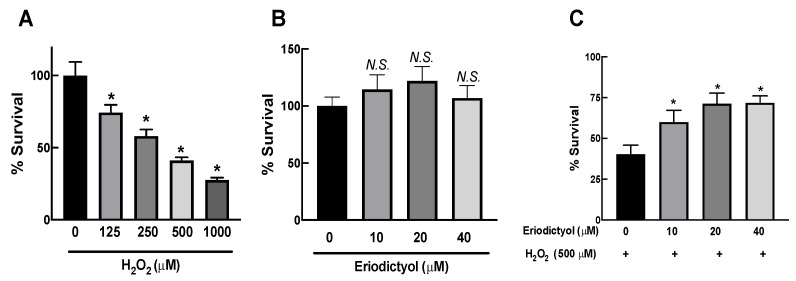
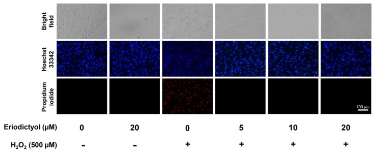
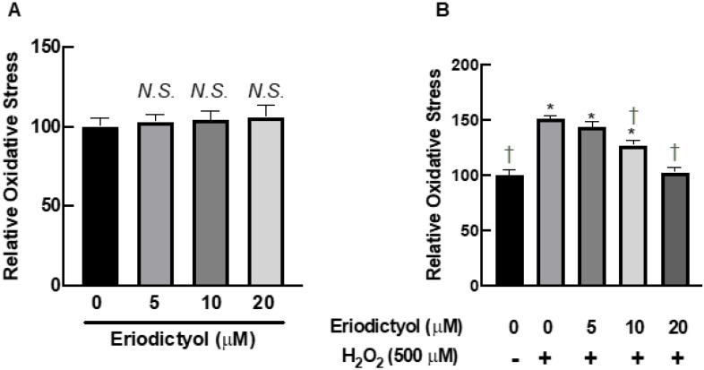
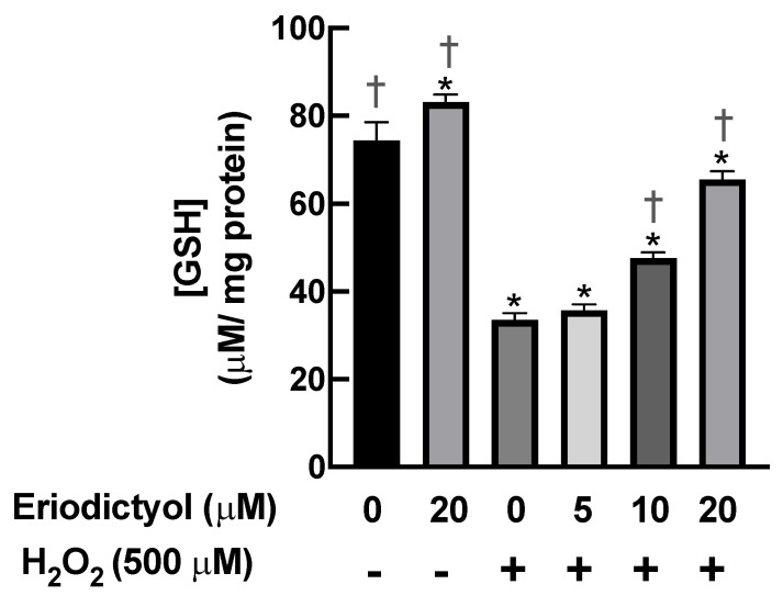
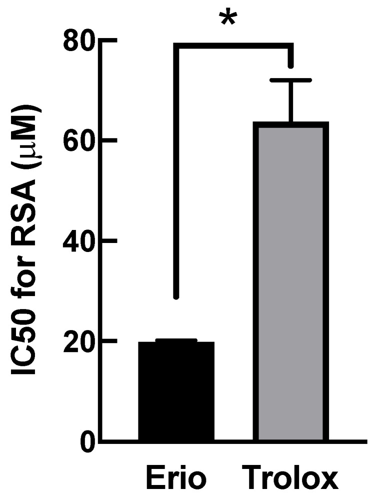
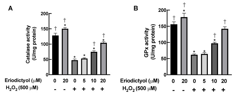
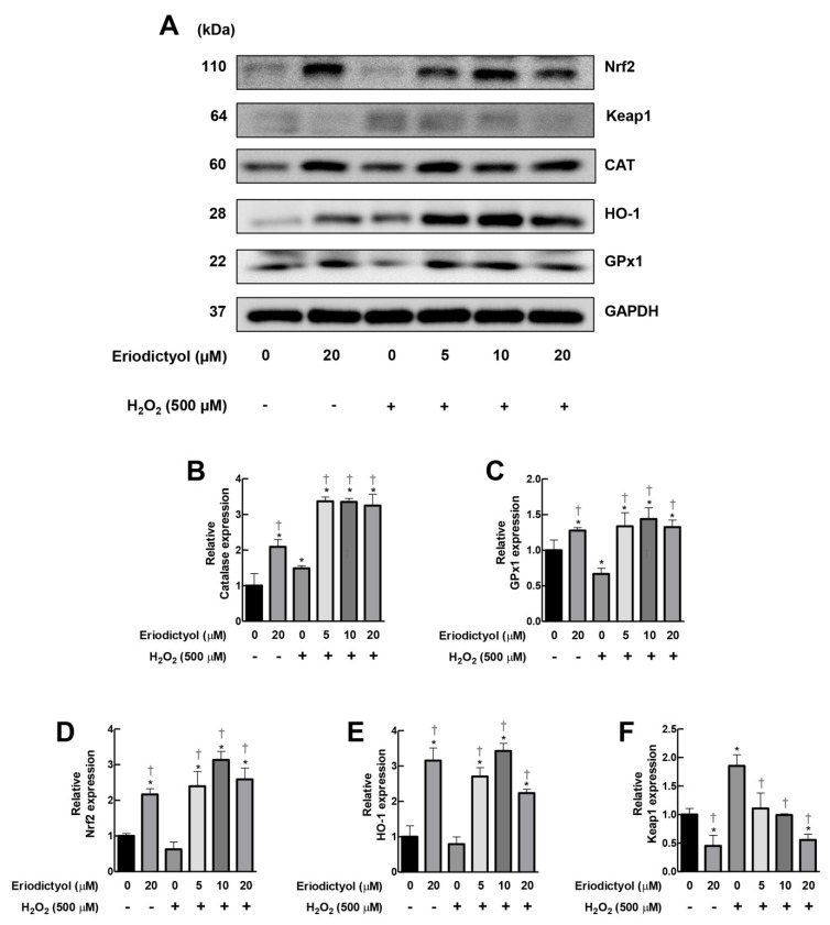
Oxidative stress in dermal fibroblasts is strongly correlated with the aging process of the skin. The application of natural compounds that can increase the ability of dermal fibroblasts to counteract oxidative stress is a promising approach to promote skin health and beauty. Eriodictyol is a flavonoid that exerts several pharmacological actions through its antioxidant properties. However, its protective effects on dermal fibroblasts have not yet been investigated. In this study, we investigated whether eriodictyol protects human dermal fibroblasts (BJ fibroblasts) from the harmful effects of hydrogen peroxide (H2O2). Eriodictyol pretreatment significantly prevented necrotic cell death caused by H2O2 exposure. In addition, the level of 2',7'-dichloro-dihydro-fluorescein oxidation was decreased, and that of glutathione was maintained, indicating that the beneficial effects of eriodictyol against H2O2 were closely associated with oxidative-stress attenuation. Eriodictyol mediates its antioxidant effects on dermal fibroblasts against H2O2 through (i) the direct neutralization of reactive oxygen species; (ii) the enhancement of the activities of H2O2-detoxifying enzymes, including catalase and glutathione peroxidase; and (iii) the induction of the expressions of catalase and glutathione peroxidase 1 via the activation of the Nrf2 signaling system. These results support the potential application of eriodictyol as an ingredient in skincare products for cosmeceutical and pharmaceutical purposes.

Keywords: antioxidants; eriodictyol; fibroblasts; hydrogen peroxide; oxidative stress; skin aging.


Conflict of interest statement

The authors declare no conflict of interest.

Figures

Figure 1
Figure 1
Eriodictyol inhibited H2O2-induced cytotoxicity in dermal fibroblasts. (A) H2O2 exposure (125–1000 µM; 1 h) resulted in a concentration-dependent decrease in the viability of BJ cells. (B) Eriodictyol treatment (10–40 µM; 24 h) did not result in cytotoxicity in BJ cells. (C) Eriodictyol pretreatment (10–40 µM; 24 h) prevented H2O2-induced cytotoxicity (500 µM; 1 h) in BJ cells. Cell viability was observed using the MTT assay after the indicated treatments. The survival percentage was calculated relative to untreated controls. DMSO (0.5%) was used as a vehicle control (n = 3; each biological replicate consisted of four independent wells; mean ± SEM). * p < 0.05 versus vehicle controls (A) and p < 0.05 versus H2O2-treated cells (C). N.S., not significant.
Figure 2
Figure 2
Eriodictyol prevented the induction of necrosis in BJ cells following H2O2 treatment. BJ cells were pre-treated with eriodictyol (5–20 µM; 24 h) followed by H2O2 treatment (500 µM; 1h). After 24 h treatment with H2O2, dermal fibroblasts were co-stained with Hoechst 33342 (blue)/PI (red) to characterize mode of cell death (n = 3; magnification = 20×; scale bar = 100 µm).
Figure 3
Figure 3
Eriodictyol inhibited oxidative stress in BJ cells following H2O2 exposure. (A) Eriodictyol treatment (5–20 µM; 24 h) did not alter the redox status of BJ cells. (B) Eriodictyol pretreatment (10–20 µM; 24 h) inhibited H2O2-induced oxidative stress (500 µM; 1 h) in a concentration-dependent fashion. Oxidative stress was immediately evaluated using the DCFH-DA assay after the indicated treatments. Relative oxidative stress was calculated compared with that in untreated controls. DMSO (0.5%) was used as a vehicle control (n = 3; each biological replicate consisted of three independent wells; mean ± SEM). * p < 0.05 versus untreated controls; p < 0.05 versus H2O2-treated cells. N.S., not significant.
Figure 4
Figure 4
Eriodictyol restored the intracellular levels of reduced glutathione following H2O2 exposure. H2O2 (500 µM; 1 h) rapidly decreased the availability of GSH in BJ cells. Eriodictyol pretreatment (10–20 µM; 24 h) maintained the intracellular levels of GSH in a concentration-dependent fashion. DMSO (0.5%) was used as a vehicle control (n = 3; each biological replicate consisted of three independent wells; mean ± SEM). * p < 0.05 versus vehicle controls; p < 0.05 versus H2O2-treated cells.
Figure 5
Figure 5
Eriodictyol has potent radical scavenging activity (RSA). The RSA of compounds was evaluated using the DPPH assay. The IC50 of eriodictyol required to decrease the radical activity of DPPH was calculated. The IC50 for the RSA of eriodictyol was 3.2-fold greater than that of Trolox (eriodictyol, 19.9 ± 0.3 µM versus Trolox, 63.8 ± 8.3 µM; n = 3; mean ± SEM, * p < 0.05).
Figure 6
Figure 6
Eriodictyol enhances the activities of CAT and GPx. (A,B) Eriodictyol pretreatment (10–20 µM; 24 h) promoted the activities of H2O2-detoxifying enzymes CAT and GPx in BJ cells following H2O2 exposure (500 µM; 1 h) in a concentration-dependent manner. DMSO (0.5%) was used as a vehicle control. Enzymatic activities were determined immediately after the treatments (n = 3; each biological replicate consisted of three independent wells; mean ± SEM). * p < 0.05 versus vehicle controls; p < 0.05 versus H2O2-treated cells.
Figure 7
Figure 7
Eriodictyol promoted the expressions of CAT and GPx1 via Nrf2 cascade. (A) Western blot assay demonstrated that supplementation with eriodictyol (5–20 µM; 24 h) enhanced the expressions of CAT and GPx1 in BJ cells following H2O2 treatment (500 µM; 1 h). The upregulation of these H2O2-removal enzymes is possibly due to the activation of the Nrf2 pathway, as observed by an increase in the expression levels of Nrf-2 and its downstream target HO-1 and a decrease in Keap1 protein expression. GAPDH was used as a loading control. DMSO (0.5%) was used as a vehicle control. Results are representative of three independent experiments. Full unedited blots are shown in Supplementary Figure S2. (BF). The band intensity of certain proteins from Western blot analysis was quantified with Image J software and normalized to loading control GAPDH. Data represent mean ± SEM of protein expression with respect to untreated control (n = 3). * p < 0.05 versus vehicle controls; p < 0.05 versus H2O2-treated cells.

References

    1. Cui H., Kong Y., Zhang H. Oxidative stress, mitochondrial dysfunction, and aging. J. Signal Transduct. 2012;2012:646354. doi: 10.1155/2012/646354. - DOI - PMC - PubMed
    1. Lephart E.D. Skin aging and oxidative stress: Equol’s anti-aging effects via biochemical and molecular mechanisms. Ageing Res. Rev. 2016;31:36–54. doi: 10.1016/j.arr.2016.08.001. - DOI - PubMed
    1. Pillai S., Oresajo C., Hayward J. Ultraviolet radiation and skin aging: Roles of reactive oxygen species, inflammation and protease activation, and strategies for prevention of inflammation-induced matrix degradation—A review. Int. J. Cosmet. Sci. 2005;27:17–34. doi: 10.1111/j.1467-2494.2004.00241.x. - DOI - PubMed
    1. Fisher G.J., Varani J., Voorhees J.J. Looking older: Fibroblast collapse and therapeutic implications. Arch. Dermatol. 2008;144:666–672. doi: 10.1001/archderm.144.5.666. - DOI - PMC - PubMed
    1. Subedi L., Lee T.H., Wahedi H.M., Baek S.-H., Kim S.Y. Resveratrol-Enriched Rice Attenuates UVB-ROS-Induced Skin Aging via Downregulation of Inflammatory Cascades. Oxidative Med. Cell. Longev. 2017;2017:8379539. doi: 10.1155/2017/8379539. - DOI - PMC - PubMed