. 2022 Jun 7;12(12):1947.
doi: 10.3390/nano12121947.

In Vitro High-Throughput Toxicological Assessment of Nanoplastics

Affiliations

In Vitro High-Throughput Toxicological Assessment of Nanoplastics

Valentina Tolardo et al. Nanomaterials (Basel). .

Abstract

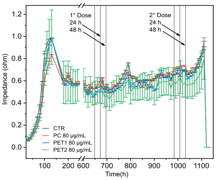
Sub-micrometer particles derived from the fragmentation of plastics in the environment can enter the food chain and reach humans, posing significant health risks. To date, there is a lack of adequate toxicological assessment of the effects of nanoplastics (NPs) in mammalian systems, particularly in humans. In this work, we evaluated the potential toxic effects of three different NPs in vitro: two NPs obtained by laser ablation (polycarbonate (PC) and polyethylene terephthalate (PET1)) and one (PET2) produced by nanoprecipitation. The physicochemical characterization of the NPs showed a smaller size, a larger size distribution, and a higher degree of surface oxidation for the particles produced by laser ablation. Toxicological evaluation performed on human cell line models (HePG2 and Caco-2) showed a higher toxic effect for the particles synthesized by laser ablation, with PC more toxic than PET. Interestingly, on differentiated Caco-2 cells, a conventional intestinal barrier model, none of the NPs produced toxic effects. This work wants to contribute to increase knowledge on the potential risks posed by NPs.

Keywords: cytotoxicity; high content screening; in vitro assays; laser ablation; nanoplastics; nanotoxicology; polycarbonate; polyethylene terephthalate.


Conflict of interest statement

The authors declare no conflict of interest.

Figures

Figure 1
Figure 1
(a) Hydrodynamic diameter (DH) distribution of PC NPs (black), PET1 NPs (red) and PET2 NPs (blue). Morphology of PC NPs (b), PET1 NPs (c) and PET2 NPs (d) by TEM. Scale bar: 100 nm.
Figure 2
Figure 2
Atomic bonding distribution from XPS C1s spectra analysis. High-resolution spectra curve-fit results from the laser ablated and PET2 NPs. Dotted lines represent stoichiometric reference values.
Figure 3
Figure 3
Mitochondrial activity of HePG2 cells after exposure to NPs at the concentrations of 1, 10, 20, 40, 80 µg/mL for 24 or 48 h (MTT assay). Data are reported as average of three independent experiments (each run in technical triplicate) ± SD. p < 0.001, p < 0.01 and p < 0.05 are reported (***, ** and * respectively), calculated versus CTRL (one-way ANOVA).
Figure 4
Figure 4
Cell viability of HePG2 and Caco-2 cells exposed to different NPs concentrations or to PC and PET NPs dispersants at 24 or 48 h. Results are expressed as percentage of cell viability compared to the untreated cells. Data are reported as the average of three independent experiments (each run in technical triplicate) ± SD. Valinomycin (900 nM) was used as positive control. p < 0.001, p < 0.01 and p < 0.05 are reported (***, ** and * respectively), calculated versus CTRL (one-way ANOVA).
Figure 5
Figure 5
Mitochondrial activity of HePG2 and Caco-2 cells exposed to different concentrations of PC or PET NPs for 24 or 48 h, measured by HCS. Results are expressed as percentage of mitochondrial activity compared to the untreated cells. Valinomycin (900 nM) was used as positive control. Data are reported as the average of three independent experiments (each run in technical triplicate) ± SD. p < 0.001, p < 0.01 and p < 0.05 are reported (***, ** and * respectively), calculated versus CTRL (one-way ANOVA).
Figure 6
Figure 6
Two-way analysis of HePG2 cells exposed to PC, PET1 or PET2 NPs at concentrations ranging between 10 and 80 μg/mL for 24 or 48 h. Valinomycin was used as positive control. Results are expressed as a function of two different parameters: mitochondrial activity and cellular membrane damage. Data are reported as the average of three independent experiments (each run in triplicate).
Figure 7
Figure 7
Two-way analysis of Caco-2 cells exposed to PC, PET1 or PET2 NPs at concentrations ranging between 10 and 80 μg/mL for 24 or 48 h. Valinomycin was used as positive control. Results are expressed as a function of two different parameters: mitochondrial activity and cellular membrane damage. Data are reported as the average of three independent experiments (each run in triplicate).
Figure 8
Figure 8
Cell viability of undifferentiated and differentiated Caco-2 cells exposed to different doses of PC, PET1 or PET2 NPs for 24 or 48 h. Data are expressed as percentage of cell viability normalized to the control (untreated cells) and reported as the mean of three independent experiments (each run in triplicate) ± SD.
Figure 9
Figure 9
Mitochondrial activity of undifferentiated and differentiated Caco-2 cells exposed to different doses of PC, PET1 or PET2 NPs for 24 or 48 h. Data are expressed as percentage of mitochondrial activity normalized to the control (untreated cells) and reported as the mean of three independent experiments (run in triplicate) ± SD.
Figure 10
Figure 10
Electrical impedance. Differentiated Caco-2 were exposed to 80 µg/mL of PC, PET1 or PET2 for 48 h; after a recovery period of 12 days, cells were re-exposed to the NPs for another 48 h. At the end of the second treatment, the medium was replaced, and cells were maintained for an additional 48 h. Triton X-100 (0.1%) was then added as positive control.

References

    1. Plastic Europe—Association of Plastics Manufactures Plastics—The Facts 2021. [(accessed on 1 June 2022)]. Available online: file:///C:/Users/MDPI/Downloads/Plastics-the-Facts-2021-web-final.pdf.
    1. Borrelle S.B., Ringma J., Lavender Law K., Monnahan C.C., Lebreton L., McGivern A., Murphy E., Jambeck J., Leonard G.H., Hilleary M.A., et al. Predicted Growth in Plastic Waste Exceeds Efforts to Mitigate Plastic Pollution. Science. 2020;369:1515–1518. doi: 10.1126/science.aba3656. - DOI - PubMed
    1. Oliveri Conti G., Ferrante M., Banni M., Favara C., Nicolosi I., Cristaldi A., Fiore M., Zuccarello P. Micro- and Nano-Plastics in Edible Fruit and Vegetables. The First Diet Risks Assessment for the General Population. Environ. Res. 2020;187:109677. doi: 10.1016/j.envres.2020.109677. - DOI - PubMed
    1. Wayman C., Niemann H. The Fate of Plastic in the Ocean Environment—A Minireview. Environ. Sci. Process. Impacts. 2021;23:198–212. doi: 10.1039/D0EM00446D. - DOI - PubMed
    1. Redondo-Hasselerharm P.E., Gort G., Peeters E.T.H.M., Koelmans A.A. Nano- and Microplastics Affect the Composition of Freshwater Benthic Communities in the Long Term. Sci. Adv. 2020;6:eaay4054. doi: 10.1126/sciadv.aay4054. - DOI - PMC - PubMed

LinkOut - more resources