. 2022 Jun 6;15(6):721.
doi: 10.3390/ph15060721.

Design and Synthesis of a Mitochondria-Targeting Radioprotectant for Promoting Skin Wound Healing Combined with Ionizing Radiation Injury

Affiliations

Design and Synthesis of a Mitochondria-Targeting Radioprotectant for Promoting Skin Wound Healing Combined with Ionizing Radiation Injury

Zaizhi Du et al. Pharmaceuticals (Basel). .

Abstract

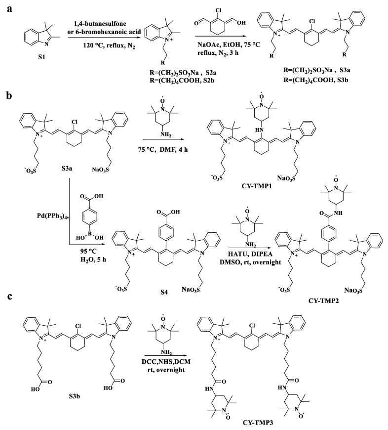
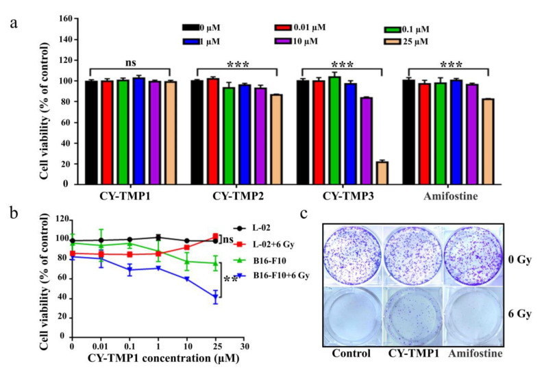
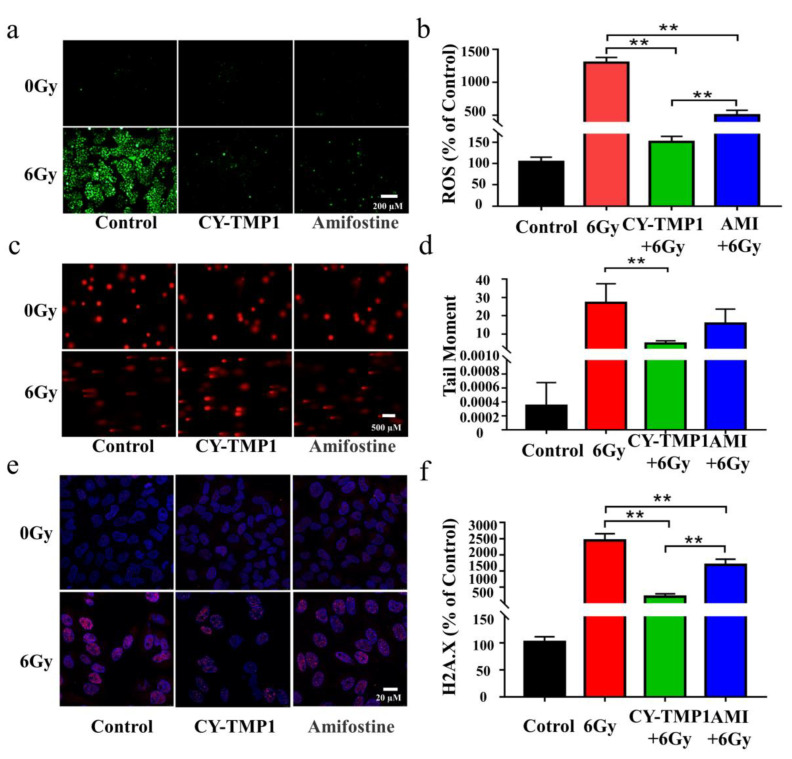
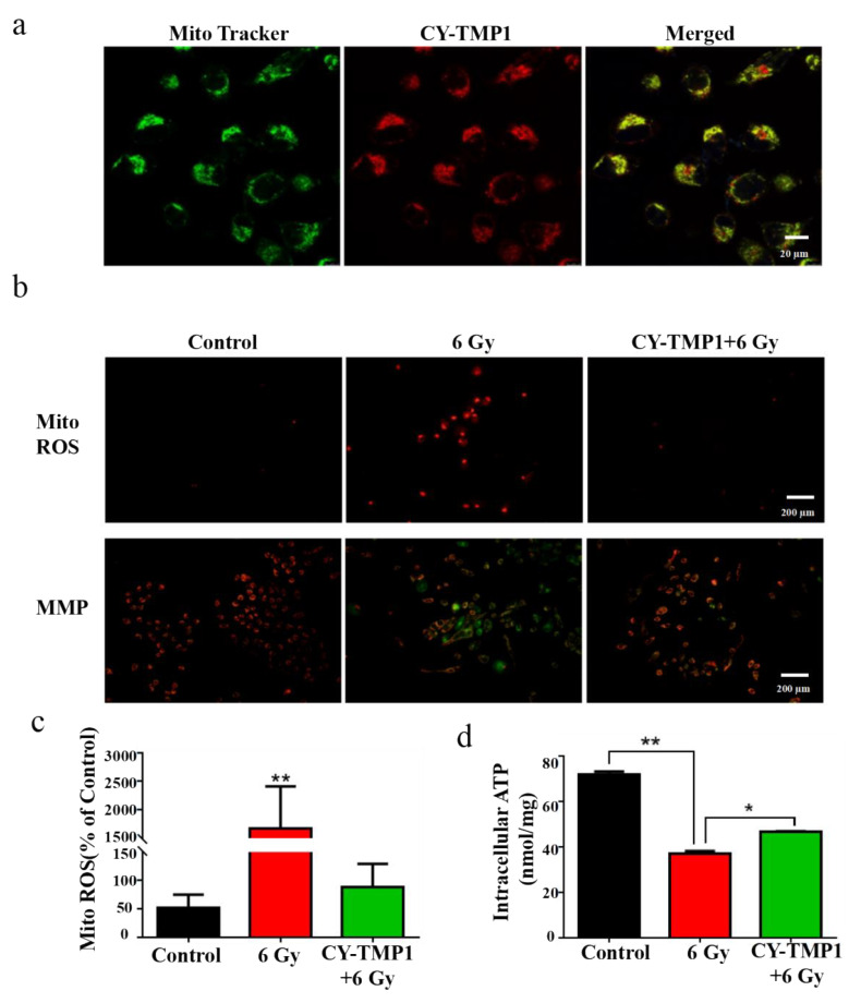
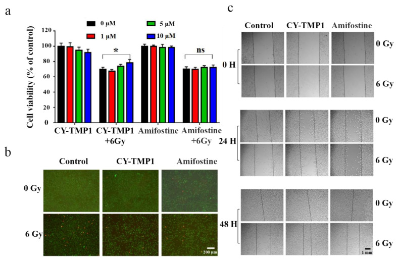
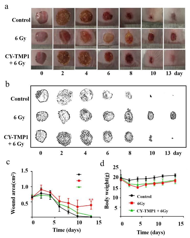
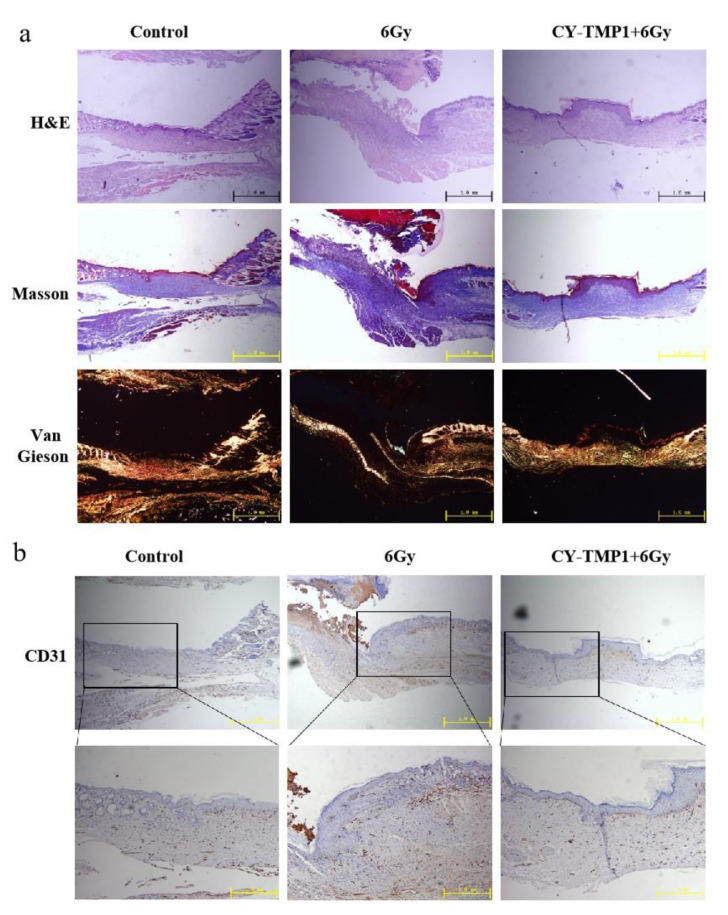
Wound healing is seriously retarded when combined with ionizing radiation injury, because radiation-induced excessive reactive oxygen species (ROS) profoundly affect cell growth and wound healing. Mitochondria play vital roles not only as cellular energy factories but also as the main source of endogenous ROS, and in this work a mitochondria-targeting radioprotectant (CY-TMP1) is reported for radiation injury-combined wound repair. It was designed, synthesized and screened out from different conjugates between mitochondria-targeting heptamethine cyanine dyes and a peroxidation inhibitor 2,2,6,6-tetramethylpiperidinyloxy (TEMPO). CY-TMP1 specifically accumulated in mitochondria, efficiently mitigated mitochondrial ROS and total intracellular ROS induced by 6 Gy of X-ray ionizing irradiation, thereby exhibiting a notable radioprotective effect. The mechanism study further demonstrated that CY-TMP1 protected mitochondria from radiation-induced injury, including maintaining mitochondrial membrane potential (MMP) and ATP generation, thereby reducing the ratio of cell apoptotic death. Particularly, an in vivo experiment showed that CY-TMP1 could effectively accelerate wound closure of mice after 6 Gy of whole-body ionizing radiation. Immunohistochemical staining further indicated that CY-TMP1 may improve wound repair through angiogenesis and re-epithelialization. Therefore, mitochondria-targeting ROS scavengers may present a feasible strategy to conquer refractory wound combined with radiation injury.

Keywords: heptamethine cyanine dye; mitochondrion; radiation protection; wound repair.


Conflict of interest statement

The authors declare no conflict of interest.

Figures

Scheme 1
Scheme 1
Synthetic routes of different TEMPO-conjugated cyanine dyes (CY-TMP). (a) Synthesis of compound S3a and S3b as the mitochondria-targeting molecular skeletons. (b) Synthesis of CY-TMP1 and CY-TMP2 by introducing TEMPO into compound S3a. (c) Synthesis of CY-TMP3 by conjugating two TEMPO groups with compound S3b.
Figure 1
Figure 1
In vitro radioprotective effects evaluation of CY-TMP conjugates. (a) Cytotoxicity evaluation of CY-TMP1, CY-TMP2, CY-TMP3 and Amifostine on L-02 cells. “***” represents significant difference from 0 μM group, “ns” represents no significance. *** p < 0.001, Data are expressed as Mean ± SD (n = 3). (b) Radioprotective effect of CY-TMP1 on normal cells (L-02) and cancer cells (B16-F10). CY-TMP1 selectively protects normal cells rather than cancer cells from ionizing radiation injury. Comparison of cell viability after treatment with CY-TMP1 (25 μM) between radiation and non-radiation groups, n = 3, ** p < 0.01, “ns” represents no significance. (c) Effect of CY-TMP1 (25 μM) on the colony formation ability of L-02 cells in the absence or presence of X-ray irradiation (6 Gy).
Figure 2
Figure 2
Comparison of radioprotective effect between CY-TMP 1 and Amifostine (Ami) on L-02 cells. (a) The total intracellular ROS induced by X-ray radiation were probed with Reactive Oxygen Species Assay Kit and (b) quantitatively measured 24 h after radiation. (c) Fluorescence imaging and (d) quantitative analysis of comet tail as an index of DNA damage. (e) Fluorescence imaging and (f) quantitative analysis of γ-H2A.X based on immunofluorescent staining. Compared to the 6 Gy radiation group, CY-TMP1 significantly decreases radiation-induced ROS level and DNA damage (n = 6, mean ± SD, ** p < 0.01). Compared to the Ami-treated group, CY-TMP1 also exhibits significantly higher efficiency in ROS-scavenging ability and reducing the ratio of DNA double chain breaks induced by ionizing radiation (n = 6, mean ± SD, ** p < 0.01).
Figure 3
Figure 3
Mechanism study of CY-TMP1 with the radioprotection effect. (a) Subcellular localization of CY-TMP1 was detected with a mitochondria-specific green probe (Mito-Tracker); (b) Alterations in mitochondrial membrane potential (MMP) and mitochondrial ROS (Mito-ROS) were detected by a fluorescence microscope before and after 6 Gy X-ray radiation; (c) Quantitative analysis of mito-ROS based on the mean fluorescent intensity of per filed with Image J software (n = 3, mean ± SD, ** p < 0.01, versus CY-TMP1+6 Gy group); (d) ATP was determined and compared with or without pretreatment of CY-TMP1, (n = 3, mean ± SD, * p < 0.05, ** p < 0.01).
Figure 4
Figure 4
Radiation protection and wound scratch assay of CY-TMP1 on HFF-1 cells. (a) Radioprotective effect of CY-TMP1 on HFF-1 cells, n = 3, * p < 0.05; “ns” represents no significance; (b) Calcein AM (green) and PI (red) probes were utilized to stain live and dead HFF-1 cells before and after 6 Gy X-ray radiation; (c) Effect of CY-TMP1 (10 μM) on the wound closure ability evaluated by a scratch assay of HFF-1 cells in the different treatment groups.
Figure 5
Figure 5
The effect of CY-TMP1 on wound repair with radiation-combined wound injury. (a) Representative photographs of wound repair at certain time points in different groups; (b) Visualization of representative photographs by Photoshop software (Photoshop CS 8.0.1, Adobe, CA, USA); (c) Quantitative measurement of wound area by Image J software (n = 6; mean ± SD, ** p < 0.01 versus 6 Gy radiation-combined wound injury group); (d) Body weight of mice in different groups. There is no statistic difference in body weight at the end of the observation (on the 13th day), indicating tolerant treatment of CY-TMP1 without apparent acute toxicity.
Figure 6
Figure 6
Histological analysis and immunofluorescence staining of wound sections at the end of observation (collected on the 13th day). (a) Representative photographs of wound sections with H&E, Masson and Van Gieson staining from three different groups; (b) Representative photographs of CD31 immunofluorescence staining of wound sections.

References

    1. Hay R.J., Johns N.E., Williams H.C., Bolliger I.W., Dellavalle R.P., Margolis D.J., Marks R., Naldi L., Weinstock M.A., Wulf S.K., et al. The global burden of skin disease in 2010: An analysis of the prevalence and impact of skin conditions. J. Investig. Dermatol. 2014;134:1527–1534. doi: 10.1038/jid.2013.446. - DOI - PubMed
    1. Devalia H.L., Mansfield L. Radiotherapy and wound healing. Int. Wound J. 2008;5:40–44. doi: 10.1111/j.1742-481X.2007.00351.x. - DOI - PMC - PubMed
    1. Gieringer M., Gosepath J., Naim R. Radiotherapy and wound healing: Principles, management and prospects (review) Oncol. Rep. 2011;26:299–307. doi: 10.3892/or.2011.1319. - DOI - PubMed
    1. Bourdais R., Achkar S., Honoré C., Faron M., Cavalcanti A., Auzac G., Ngo C., Haddag-Miliani L., Verret B., Dumont S., et al. Prospective evaluation of intensity-modulated radiotherapy toxicity in extremity soft tissue sarcomas patients: A role for irradiated healthy soft tissue volume? Clin. Transl. Radiat. Oncol. 2021;29:79–84. doi: 10.1016/j.ctro.2021.05.007. - DOI - PMC - PubMed
    1. Anderson K., Hamm R.L. Factors That Impair Wound Healing. J. Am. Coll. Clin. Wound Spec. 2012;4:84–91. doi: 10.1016/j.jccw.2014.03.001. - DOI - PMC - PubMed

LinkOut - more resources