. 2022 May 27;14(6):1137.
doi: 10.3390/pharmaceutics14061137.

Characterization of Physical and Biological Properties of a Caries-Arresting Liquid Containing Copper Doped Bioglass Nanoparticles

Affiliations

Characterization of Physical and Biological Properties of a Caries-Arresting Liquid Containing Copper Doped Bioglass Nanoparticles

Se-Jung Bang et al. Pharmaceutics. .

Abstract

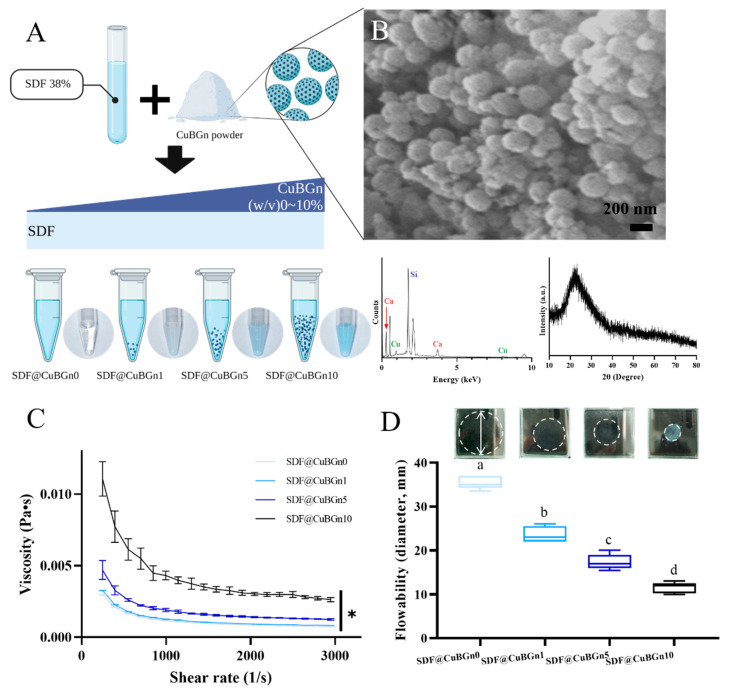
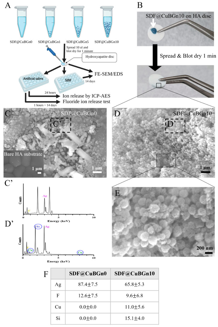
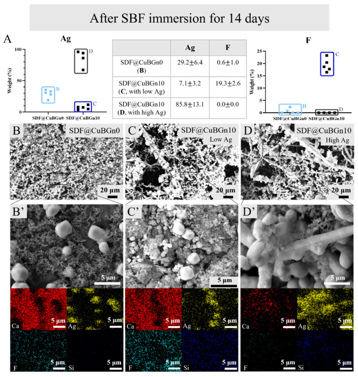
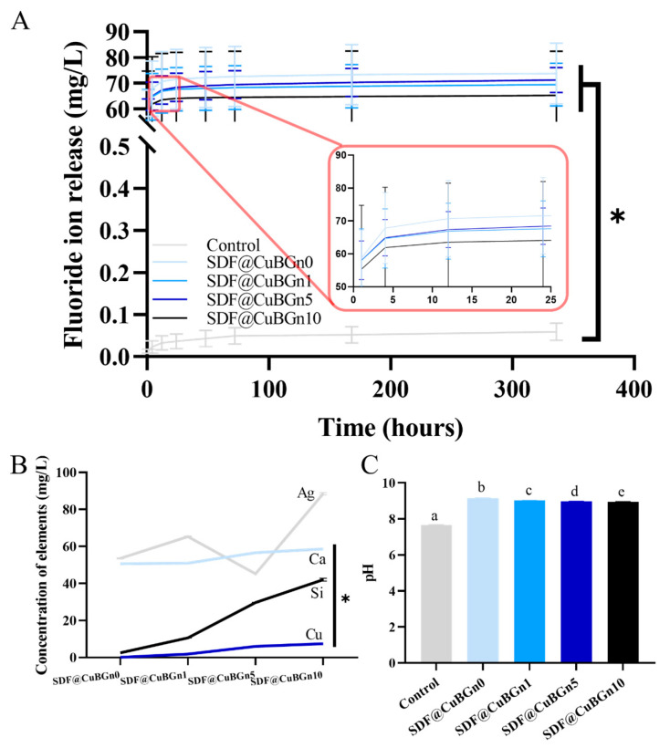
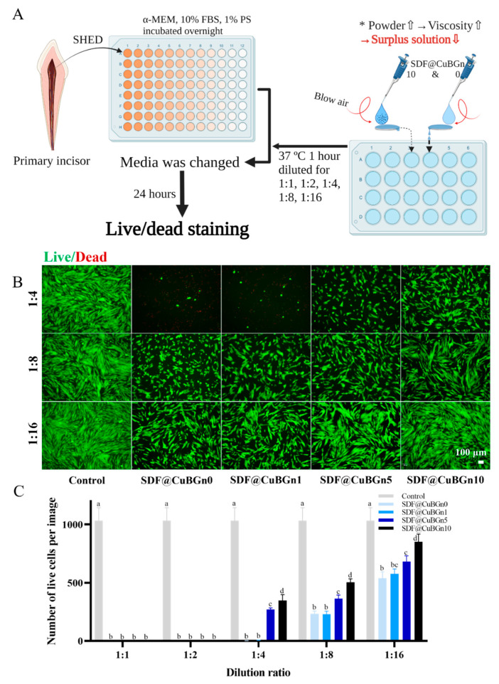
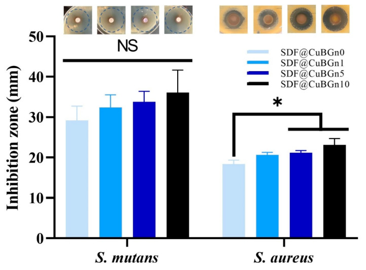
Silver diamine fluoride (SDF) is an outstanding dental material for arresting and preventing caries, but some drawbacks, such as high flowability due to low viscosity and cytotoxicity to the pulp, have been reported. To overcome these problems, copper-doped bioactive glass nanoparticles (CuBGns) were combined with SDF. After synthesis, CuBGns were examined by physical analysis and added in SDF at different weight/volume% (SDF@CuBGn). After assessing physical properties (viscosity and flowability) of SDF@CuBGn, physicochemical properties (morphology before and after simulated body fluid (SBF) immersion and ion release) of SDF@CuBGn-applied hydroxyapatite (HA) discs were evaluated. Biological properties were further evaluated by cytotoxicity test to pulp stem cells and antibacterial effect on cariogenic organisms (Streptococcus mutans and Staphylococcus aureus). Combining CuBGns in SDF increased the viscosity up to 3 times while lowering the flowability. More CuBGns and functional elements in SDF (Ag and F) were deposited on the HA substrate, even after SBF immersion test for 14 days, and they showed higher Cu, Ca, and Si release without changing F and Ag release. Cell viability test suggested lower cytotoxicity in SDF@CuBGn-applied HA, while CuBGns in SDF boosted antibacterial effect against S. aureus, ~27% in diameter of agar diffusion test. In conclusion, the addition of CuBGn to SDF enhances viscosity, Ag and F deposition, and antibacterial effects while reducing cell toxicity, highlighting the role of bioactive CuBGns for regulating physical and biological effects of dental materials.

Keywords: Staphylococcus aureus; Streptococcus mutans; biological properties; copper-doped bioactive glass nanoparticle; hydroxyapatite disc; physicochemical properties; pulp stem cells; silver diamine fluoride; viscosity.


Conflict of interest statement

The authors declare no conflict of interest.

Figures

Figure 1
Figure 1
Preparation and characterization of copper-doped bioactive glass nanoparticles (CuBGns) and physical analysis of a silver diamine fluoride (SDF) solution mixed with CuBGns (SDF@CuBGns). (A) Schematic illustration of the preparation of SDF@CuBGn and a picture of the actual mix. CuBGn was placed in SDF for 0, 1, 5, and 10 (w/v)%, and the larger the amount of powder was, the more blue the solution. (B) Field emission scanning electron microscopy (FE-SEM) (50,000×, 5 kV), energy dispersive spectroscopy (EDS) and X-ray diffraction (XRD) of CuBGns. FE-SEM shows the numerous spherical morphologies of CuBGns, and EDS analysis confirmed the presence of elements Si, Cu and Ca. The presence of a broad halo (at 2θ = 20° to 30°) and absence of XRD diffraction peaks showed the amorphous structure. (C) Viscosity and (D) flowability of SDF@CuBGn. Viscosity was measured with 0.3 mL of sample placed between parallel plates (diameter 60.0 mm) (n = 3). The flow test was performed according to ISO 6876:2012. Then, 0.01 mL of SDF@CuBGn was placed onto a 40 mm × 40 mm, 5 mm thick glass plate (approximately 20 g) and covered with an identical glass plate. An additional 100 g of weight was placed on the sample and removed after 10 min. The maximum and minimum diameters of SDF@CuBGn were measured and averaged (n = 5). Adding more CuBGn resulted in a higher viscosity and lower flowability. There was a significant difference between groups with different letters (a, b, c, and d) in (D). Statistical analysis was performed using one-way analysis of variance (ANOVA) followed by Tukey’s post-hoc test. * p < 0.05.
Figure 2
Figure 2
Physicochemical analysis (morphology and surface elemental analysis) of hydroxyapatite (HA) discs with SDF@CuBGn. (A) Schematic illustration of the physicochemical analysis of HA discs with SDF@CuBGn. Ten microliters of SDF@CuBGn were spread on HA discs (diameter 9.5 mm, thickness 1.8 mm) and blotted for 1 min. Discs were prepared and examined by FE-SEM and EDS after SDF@CuBGn was applied, immersed in simulated body fluid (SBF, at pH 7.4 and 37 °C) for 14 days, and ion release tests were performed after soaking in artificial saliva (37 °C, being agitated). (B) Actual picture of spreading SDF@CuBGn10 on HA discs. (C) HA disc with no treatment (bottom left) and SDF@CuBGn0, (D) SDF@CuBGn10 on HA disc analyzed with FE-SEM (10,000×, 20 kV) and (C’), (D’) EDS analysis of parts of (C) and (D). Ag and F were found in both samples, and Cu and Si were detected only in SDF@CuBGn10. (E) Magnified FE-SEM of SDF@CuBGn10 (40,000×, 20 kV). The spherical morphology of CuBGn, which was not seen in (C) SDF@CuBGn0, was found. (F) EDS analysis of components of SDF@CuBGn-applied HA discs (weight%).
Figure 3
Figure 3
Physicochemical analysis (surface analysis after SBF immersion for 14 days) of SDF@CuBGn-applied HA discs. (A) Elemental EDS analysis of Ag and F. (B) SDF@CuBGn0-applied HA disc and (B’) magnified image. (C,D) SDF@CuBGn10-applied HA disc and (C’,D’) magnified image. SDF@CuBGn0- and SDF@CuBGn10-applied HA discs were immersed in SBF, which is optimal condition for accelerating remineralization. After 14 days, the samples were thoroughly washed, and the morphological change was observed by FE-SEM and EDS (200×, 5000×, 15 kV). SDF@CuBGn-applied discs showed many block-shape mass and EDS analysis revealed that they were Ag. Particularly, SDF@CuBGn10-applied disc showed that the silver blocks held together to form rod-shaped branches of Ag. The spot with lots of silver rods (D) showed very high silver content, while the other spots (C) showed high fluoride deposition. Adding CuBGns made silver-branch and even increased fluoride deposition in spots with low silver.
Figure 4
Figure 4
Physicochemical analysis (ion release tests for F, Ca, Cu, and Si and pH measurements) of SDF@CuBGn-applied HA discs. (A) Accumulated fluoride ion release test. More than 90% of fluoride was released within 4 h, and all test groups presented significantly higher (at least 1000 times) fluoride ion release than the control group (p < 0.05). CuBGn did not affect the result. (p > 0.05). (B) Ag, Ca, Cu, and Si ion concentrations and (C) pH measurements of artificial saliva with SDF@CuBGn-applied discs in it for 24 h. As the CuBGn increased, the concentration of released Ca, Cu, and Si ions also increased (p < 0.05) without affecting Ag ion release (p > 0.05), and the pH of the extracted media decreased, although it was higher than that of the control solution. There was a significant difference between groups with different letters (a, b, c, d, and e) in (C).Interestingly, adding CuBGn slightly decreased the pH despite hydrolysis of BGn producing OH ions, which may have been due to the amount of SDF@CuBGn attached to the HA disc increasing as the viscosity increased. Statistical analysis was performed using ANOVA and repeated-measures ANOVA only in the fluoride release test followed by Tukey’s post-hoc test. * p < 0.05, n = 3.
Figure 5
Figure 5
In vitro study of cytotoxicity on stem cells from human exfoliated deciduous teeth (SHED). (A) Schematic illustration of the cytotoxicity test of SDF@CuBGn on SHED. The higher the viscosity, the more solution was expected to attach to the HA disc. Based on this, the test was designed to simulate when SDF@CuBGn was applied to teeth. Ten microliters of SDF@CuBGn were placed on one end of the HA disc, and compressed air was blown to flow the solution to the other side. The surplus solution that fell into α-minimum essential medium (α-MEM) was used for the SHED cell viability test, and as expected, the relatively high-viscosity solution had less surplus solution. (B) Fluorescence image of live (green color)/dead (red color) cells. (C) Number of live cells per image. Surplus test solutions were diluted to 1:1, 1:2, 1:4, 1:8, and 1:16. At 1:1 and 1:2 dilution ratios in culture media, all groups of cells did not survive. At a 1:4 dilution ratio, SDF@CuBGn5 and SDF@CuBGn10 showed lower cell viability (approximately 30% compared to the control group), but both SDF@CuBGn0 and SDF@CuBGn1 had extremely low cell viability (<1% compared to the control group). At all dilution ratios, as the amount of powder increased, cell viability also increased due to the diminished surplus solution. Statistical analysis was performed using ANOVA followed by Tukey’s post-hoc test. There was a significant difference between groups with different letters (a, b, c, and d) in (C). * p < 0.05, n = 6.
Figure 6
Figure 6
In vitro study of antibacterial effects on cariogenic organisms. To measure the antibacterial effect, agar diffusion tests were selected, which are simple, fast, and reliable. In Streptococcus mutans (S. mutans) and Staphylococcus aureus (S. aureus), the diameter of the inhibition zone increased as the powder increased, but it was not statistically significant in S. mutans (p > 0.05, expressed as NS). In S. aureus, SDF@CuBGn5 and SDF@CuBGn 10 clearly had greater effects than SDF@CuBGn0 (p < 0.05). Silver ions exhibit antibacterial effects, and in addition, CuBGn releases copper ions even under bacterial infection and has antimicrobial activity for hard tissue repair, so it was expected to show higher antimicrobial activity when it was combined with SDF. Statistical analysis was performed using ANOVA followed by Tukey’s post-hoc test. * p < 0.05, n = 3.

References

    1. Tinanoff N., Douglass J.M. Clinical decision-making for caries management in primary teeth. J. Dent. Educ. 2001;65:1133–1142. doi: 10.1002/j.0022-0337.2001.65.10.tb03462.x. - DOI - PubMed
    1. Sheiham A. Impact of dental treatment on the incidence of dental caries in children and adults. Community Dent. Oral Epidemiol. 1997;25:104–112. doi: 10.1111/j.1600-0528.1997.tb00906.x. - DOI - PubMed
    1. Pitts N.B., Zero D.T., Marsh P.D., Ekstrand K., Weintraub J.A., Ramos-Gomez F., Tagami J., Twetman S., Tsakos G., Ismail A. Dental caries. Nat. Rev. Dis. Primers. 2017;3:1–16. doi: 10.1038/nrdp.2017.30. - DOI - PubMed
    1. Peng J.-Y., Botelho M., Matinlinna J. Silver compounds used in dentistry for caries management: A review. J. Dent. 2012;40:531–541. doi: 10.1016/j.jdent.2012.03.009. - DOI - PubMed
    1. Rosenblatt A., Stamford T., Niederman R. Silver diamine fluoride: A caries “silver-fluoride bullet”. J. Dent. Res. 2009;88:116–125. doi: 10.1177/0022034508329406. - DOI - PubMed

LinkOut - more resources