. 2022 Jun 13;14(6):1257.
doi: 10.3390/pharmaceutics14061257.

HO-3867 Induces Apoptosis via the JNK Signaling Pathway in Human Osteosarcoma Cells

Affiliations

HO-3867 Induces Apoptosis via the JNK Signaling Pathway in Human Osteosarcoma Cells

Peace Wun-Ang Lu et al. Pharmaceutics. .

Abstract

Metastatic osteosarcoma often results in poor prognosis despite the application of surgical en bloc excision along with chemotherapy. HO-3867 is a curcumin analog that induces cell apoptosis in several cancers, but the apoptotic effect and its mechanisms on osteosarcoma cells are still unknown. After observing the decrease in cellular viability of three human osteosarcoma U2OS, HOS, and MG-63 cell lines, and the induction of cellular apoptosis and arrest in sub-G1 phase in U2OS and HOS cells by HO-3867, the human apoptosis array showed that heme oxygenase (HO)-1 and cleaved caspase-3 expressions had significant increases after HO-3867 treatment in U2OS cells and vice versa for cellular inhibitors of apoptosis (cIAP)1 and X-chromosome-linked IAP (XIAP). Western blot analysis verified the results and showed that HO-3867 activated the initiators of both extrinsic caspase 8 and intrinsic caspase 9, and significantly increased cleaved PARP expression in U2OS and HOS cells. Moreover, with the addition of HO-3867, ERK1/2, and JNK1/2 phosphorylation were increased in U2OS and HOS cells. Using the inhibitor of JNK (JNK in 8), HO-3867's increases in cleaved caspases 3, 8, and 9 could be expectedly suppressed, indicating that JNK signaling is responsible for both apoptotic pathways, including extrinsic and intrinsic, in U2OS and HOS cells caused by HO-3867. Through JNK signaling, HO-3867 has proven to be effective in causing both extrinsic and intrinsic apoptotic pathways of human osteosarcoma cells.

Keywords: ERK; HO-3867; JNK; apoptosis; curcumin; osteosarcoma.


Conflict of interest statement

The authors declare no conflict of interest.

Figures

Figure 1
Figure 1
Analysis of cell viability in free and HO-3867-treated U2OS, HOS and MG-63 cells. (A) The chemical structures of curcumin and curcumin analog HO-3867 were drawn; (B) MTT assay was employed to detect the viability of U2OS, HOS, and MG-63 cells, which were treated with HO-3867 (2, 4, 8, 16, and 32 μM) for 24 h. After quantitative analysis, the effects are illustrated. n = 4. U2OS: F = 913.460, p < 0.001; HOS: F = 1110.912, p < 0.001; MG-63 (n ≥ 4): F = 880.549, p < 0.001; (C,D) CCk-8 assay was employed to detect the viability of U2OS and HOS cells, which were treated with HO-3867 (2, 4, 8, 16, and 32 μM) for 24 h. After quantitative analysis, the effects are illustrated. U2OS: F = 270.171, p < 0.001; HOS: F = 390.389, p < 0.001; a p < 0.05, when compared to 0 μM. b p < 0.05, when compared to 2 μM. c p < 0.05, when compared to 4 μM. d p < 0.05, when compared to 8 μM. e p < 0.05, when compared to 16 μM.
Figure 2
Figure 2
Analysis of cell cycle in HO-3867 treated U2OS and HOS cells. (A) After treatment with experimental concentration range of HO-3867 for 24 h, flow cytometry after PI was performed to determine DNA contents in U2OS and HOS cells; (B,C) The cell cycle profile of flow cytometry was subsequently quantified.
Figure 3
Figure 3
Analysis of cell apoptosis in HO-3867-treated U2OS and HOS cells. (A) After treatment with experimental concentration range of HO-3867 for 24 h, Annexin V-FITC/PI staining was performed to analyze DNA contents in U2OS and HOS cells. (B,C) The Annexin V-positive cells were subsequently quantified. U2OS: n = 6, F = 355.589, p < 0.001; HOS: n = 5, F = 18.887, p < 0.001. a Significantly different, p < 0.05, when compared to control. b Significantly different, p < 0.05, when compared to 2 μM. c Significantly different, p < 0.05, when compared to 4 μM. d Significantly different, p < 0.05, when compared to 8 μM.
Figure 4
Figure 4
Analysis of the human apoptosis array and IAPs in HO-3867 treated U2OS cells. (A) After treatment with 8 μM of HO-3867 for 24 h in U2OS cells, the human apoptosis array was employed. The increased proteins cleaved caspase-3 and HO-1 as well as the decreased proteins cIAP-1 and XIAP were subjected to quantitative analysis; (B) After experimental concentration range of HO-3867 treatment for 24 h, Western blot analysis was performed to measure expressions of HO-1, cIAP-1, and XIAP in U2OS and HOS cells. Then, quantitative analysis was assessed. n = 3. HO-1: U2OS: F = 601.182, p < 0.001; HOS: F = 3705.167, p < 0.001; cIAP-1: U2OS: F = 227.563, p < 0.001; HOS: F = 864.074, p < 0.001; XIAP: F = 227.563, p < 0.001; HOS: F = 864.074, p < 0.001. a Significantly different, p < 0.05, when compared to control. b Significantly different, p < 0.05, when compared to 2 μM. c Significantly different, p < 0.05, when compared to 4 μM. d Significantly different, p < 0.05, when compared to 8 μM.
Figure 5
Figure 5
Analysis of activation of caspases 8, 9, and 3 in free and HO-3867-treated U2OS and HOS cells. After experimental concentration range of HO-3867 treatment for 24 h, Western blot analysis was performed to measure expressions of caspases 8, 9, and 3, and PARP as well as their active forms in (A,B) U2OS and (C,D) HOS cells. Then, quantitative analysis was assessed. n = 3. Caspase 8: U2OS: F = 4689.887, p < 0.001; HOS: F = 310.316, p < 0.001; caspase 9: U2OS: F = 1818.961, p < 0.001; HOS: F = 212.948, p < 0.001; caspase 3: U2OS: F = 7609.925, p < 0.001; HOS: F = 1718.653, p < 0.001; PARP: U2OS: F = 406.910, p < 0.001; HOS: F = 528.206, p < 0.001. Cleaved caspase 8: U2OS: F = 326.549, p = 0.01; HOS: F = 689.730, p < 0.001; cleaved caspase 9: U2OS: F = 36.155, p < 0.001; HOS: F = 96.503, p < 0.001; cleaved caspase 3: U2OS: F = 9138.156, p < 0.001; HOS: F = 10,315.365, p < 0.001; cleaved PARP: U2OS: F = 729.355, p < 0.001; HOS: F = 405.954, p < 0.001. a Significantly different, p < 0.05, when compared to control. b Significantly different, p < 0.05, when compared to 2 μM. c Significantly different, p < 0.05, when compared to 4 μM. d Significantly different, p < 0.05, when compared to 8 μM.
Figure 6
Figure 6
Analysis of phosphorylation of ERK1/2, JNK1/2, and p38 in HO-3867-treated cells. (A,B) After experimental concentration range of HO-3867 treatment for 24 h, Western blot analysis was performed to measure expressions of MAPKs, as well as their phosphorylation in (A) U2OS and (B) HOS cells. (C,D) Next, quantitative analysis was assessed. n = 3. p-ERK: U2OS: F = 661.501, p < 0.001; HOS: F = 4585.730, p < 0.001; p-JNK: U2OS: F = 494.446, p < 0.001; HOS: F = 855.033, p < 0.001; p-p38: U2OS: F = 33.591, p < 0.001; HOS: F = 23.845, p < 0.001. a Significantly different, p < 0.05, when compared to control. b Significantly different, p < 0.05, when compared to 2 μM. c Significantly different, p < 0.05, when compared to 4 μM. d Significantly different, p < 0.05, when compared to 8 μM.
Figure 7
Figure 7
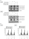
Analysis of HO-3867 on cleaved caspases 8, 9 and 3 expressions in HO-3867 with or without inhibitors of ERK1/2 (U0126)- and JNK1/2 (JNK-IN-8)-treated U2OS and HOS cells. (A) After pretreatment with or without 10 μM of U0126 and 1 μM of JNK-IN-8 for 1 h followed by 8 μM or no HO-3867 treatment for an additional 24 h, Western blot analysis was performed to measure expressions of cleaved caspases 3, 8, and 9 in U2OS and HOS cells; (B) Subsequently, quantitative analysis was assessed. n = 3. Cleaved caspase 8: U2OS: F = 536.028, p < 0.001; HOS: F = 719.244, p < 0.001; cleaved caspase 9: F = 550.879, p < 0.001; HOS: F = 436.343, p < 0.001; cleaved caspase 3: U2OS: F = 693.877, p < 0.001; HOS: F = 2338.671, p < 0.001. a Significantly different, p < 0.05, when compared to control. b Significantly different, p < 0.05, when compared to 8 μM HO-3867. c Significantly different, p < 0.05, when compared to U0126. d Significantly different, p < 0.05, when compared to U0126 and HO-3867 treatment. e Significantly different, p < 0.05, when compared to JNK-IN-8.

References

    1. Mirabello L., Troisi R.J., Savage S.A. Osteosarcoma incidence and survival rates from 1973 to 2004: Data from the surveillance, epidemiology, and end results program. Cancer. 2009;115:1531–1543. doi: 10.1002/cncr.24121. - DOI - PMC - PubMed
    1. Ottaviani G., Jaffe N. The epidemiology of osteosarcoma. Cancer Treat. Res. 2009;152:3–13. - PubMed
    1. Arndt C.A., Crist W.M. Common musculoskeletal tumors of childhood and adolescence. N. Engl. J. Med. 1999;341:342–352. doi: 10.1056/NEJM199907293410507. - DOI - PubMed
    1. Lu K.H., Lu E.W., Lin C.W., Yang J.S., Yang S.F. New insights into molecular and cellular mechanisms of zoledronate in human osteosarcoma. Pharmacol. Ther. 2020;214:107611. doi: 10.1016/j.pharmthera.2020.107611. - DOI - PubMed
    1. Oertel S., Blattmann C., Rieken S., Jensen A., Combs S.E., Huber P.E., Bischof M., Kulozik A., Debus J., Schulz-Ertner D. Radiotherapy in the treatment of primary osteosarcoma—A single center experience. Tumori. 2010;96:582–588. doi: 10.1177/030089161009600411. - DOI - PubMed