Review
. 2022 Jun 16;14(6):1283.
doi: 10.3390/pharmaceutics14061283.

A Historical Review of Brain Drug Delivery

Affiliations
Review

A Historical Review of Brain Drug Delivery

William M Pardridge. Pharmaceutics. .

Abstract

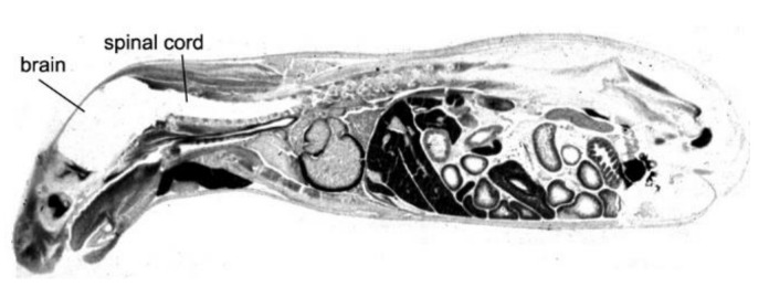
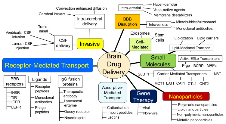
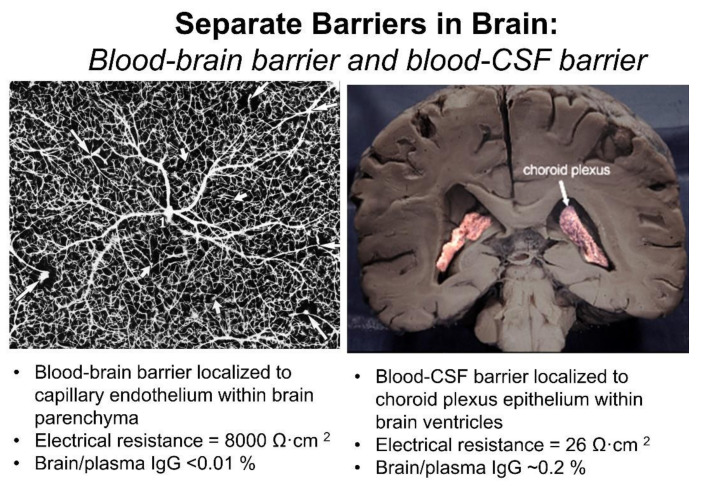
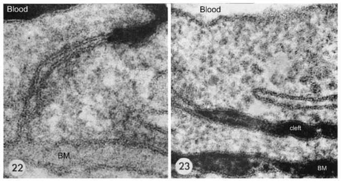
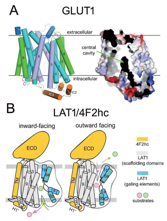
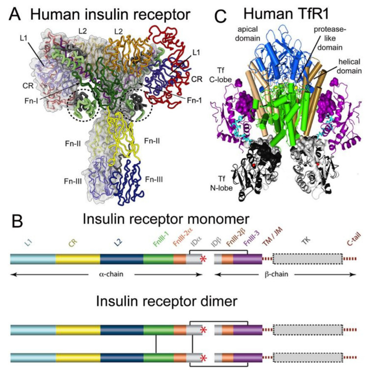
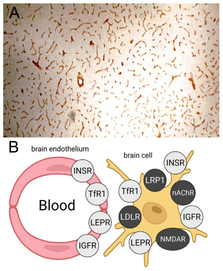
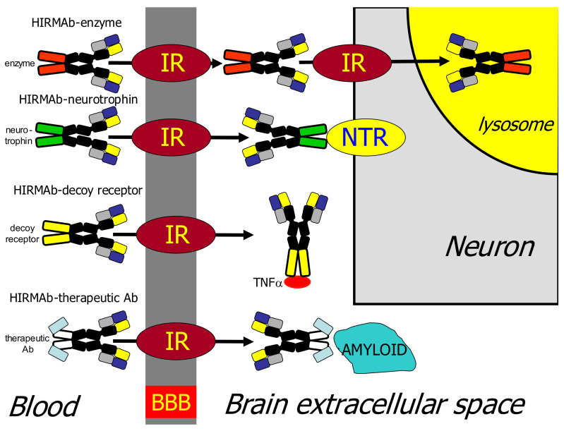
The history of brain drug delivery is reviewed beginning with the first demonstration, in 1914, that a drug for syphilis, salvarsan, did not enter the brain, due to the presence of a blood-brain barrier (BBB). Owing to restricted transport across the BBB, FDA-approved drugs for the CNS have been generally limited to lipid-soluble small molecules. Drugs that do not cross the BBB can be re-engineered for transport on endogenous BBB carrier-mediated transport and receptor-mediated transport systems, which were identified during the 1970s-1980s. By the 1990s, a multitude of brain drug delivery technologies emerged, including trans-cranial delivery, CSF delivery, BBB disruption, lipid carriers, prodrugs, stem cells, exosomes, nanoparticles, gene therapy, and biologics. The advantages and limitations of each of these brain drug delivery technologies are critically reviewed.

Keywords: IgG fusion proteins; blood–brain barrier; carrier-mediated transport; endothelium; genetic engineering; liposomes; nanoparticles; receptor-mediated transport.


Conflict of interest statement

W.M.P. is an inventor of patents on the delivery of biologics to brain.

Figures

Figure 1
Figure 1
The blood–brain barrier to small molecules. Whole body autoradiography of mouse following the IV injection of [14C]-histamine shows the lack of transport of this small molecule drug into brain and spinal cord. Reprinted with permission from [2], Copyright© 1986 American College of Physicians.
Figure 2
Figure 2
Overview of brain drug delivery technologies. These multiple delivery technologies can be broadly classified into 3 categories: (i) invasive brain drug delivery, which includes trans-cranial intra-thecal drug delivery into CSF, intra-cerebral implants, or convection-enhanced diffusion; (ii) BBB disruption brain drug delivery, which includes either the intra-carotid arterial infusion of noxious agents, or the intravenous injection of microbubbles coupled with focal external sonication of the brain; (iii) trans-vascular brain drug delivery, which includes receptor-mediated transport, carrier-mediated transport, active efflux transport, and lipid-mediated transport.
Figure 3
Figure 3
Blood–brain barrier vs. blood–CSF barrier. (Left) Inverted India ink labeling of microvasculature of human cerebral cortex, which is from [14] with permission, Copyright© 1981 Elsevier. (Right) Coronal section of human brain showing the choroid plexus lining the floor of both lateral ventricles. Adapted from [15], Copyright© 2020 licensed under Creative Commons Attribution License (CC-BY).
Figure 4
Figure 4
Brain capillary endothelial tight junctions. (Left) Electron microscopy of mouse brain following the intravenous injection of lanthanum, which is retained in the blood volume of brain at the top of the figure. (Right) Electron microscopic histochemistry of mouse brain following the intrathecal injection of HRP. This 40 kDa protein moves freely through the brain ECS, through the astrocyte endfeet, through the capillary basement membrane (BM), and into abluminal clefts formed between adjacent endothelial cells. Reproduced from [3], Copyright© 1969 under Creative Commons Attribution License (Share Alike 4.0 Unported).
Figure 5
Figure 5
Limited drug delivery to brain via the ventricular CSF. (A) Peroxidase histochemistry of mouse brain removed at either 10 min (left) or 90 min (right) after ICV administration of HRP. The magnification bar is 0.7 mm. Reproduced from [3], Copyright© 1969 under Creative Commons Attribution License (Share Alike 4.0 Unported). (B) Brain concentrations of hydroxyurea (MW = 76 Da), methotrexate (MW = 454 Da), and thiotepa (MW = 189 Da) at 1–4 mm, removed from ependymal surface at 60 min following drug injection into the lateral ventricle of the Rhesus monkey. Reproduced with permission from [78], Copyright© 1975 Am. Soc. Pharm. Exp. Ther. (C) Film autoradiography of a coronal section of rat brain removed 24 h after injection into one lateral ventricle (LV) of [125I]-BDNF. The magnification bar is 2 mm; 3V = third ventricle. Reproduced with permission from [79], Copyright© 1994 Elsevier.
Figure 6
Figure 6
Brain drug delivery by convection-enhanced diffusion. (A) Concentration of GDNF in primate brain at 1–7 mm removed from the CED catheter. Derived from data reported by Salvatore et al. [131] and reproduced with permission from [132], Copyright© 2010 Taylor and Francis. Original GDNF concentrations, in pg per mg protein, were converted to ng per gram brain, based on 100 mg protein per gram brain [133]. (B,C) Glial fibrillary acidic protein (GFAP) immunohistochemistry of monkey brain following CED administration of GDNF. Magnification bar is 1 mm in (B) and 50 microns in (C). Reproduced with permission [134], Copyright© 2003 John Wiley & Sons.
Figure 7
Figure 7
Anatomy of the cribriform plate separation of the cranial and nasal cavities. (A) The bipolar olfactory sensory neurons, which express the olfactory receptors, pass from the olfactory bulb to the nasal mucosa through the fenestrations of the cribriform plate. Reproduced with permission from [149], Copyright© 2003 JNS Publishing Group. (B) Magnetic resonance imaging (MRI) of the human head between 3–6 h following the injection into the lumbar CSF of gadobutrol, a macrocyclic gadolinium contrast agent. The gadolinium is visualized throughout the cerebral subarachnoid space within the convexities of the cerebrum, around the spinal cord, and is observed to penetrate into the superior regions of the fenestrations of the cribriform plate, which is magnified in the inset. No gadolinium passes into the inferior regions of the cribriform fenestrations, or into the nasal mucosa in humans (inset). Reproduced from [150], Copyright© 2020 licensed under Creative Commons Attribution License (CC-BY).
Figure 8
Figure 8
Overton vs. Stein models of solute diffusion through membranes. (A) Overton model of solute diffusion through biological membranes. Membrane permeability is independent of solute molecular size [315]. (B) Stein model of solute diffusion through membranes [317]. Membrane permeability is exponentially related to the molecular volume of the drug (Vd) relative to the volume (Vh) of transitory holes formed in the membrane. These membrane holes are formed by kinking of phospholipid fatty acyl side chains, as depicted in the model. Adapted from [15], Copyright© 2020 licensed under Creative Commons Attribution License (CC-BY).
Figure 9
Figure 9
Structure of GLUT1 and LAT1 carrier-mediated transporters. (A) (Left) Model of crystal structure of human GLUT1 showing orientation of 12 transmembrane regions (TMR) in four 3-helical repeat domains composed of TMRs 1,4,7,10 (blue), TMRs 3,6,9,12 (green), and TMRs 2,5,8,11 (purple); the extracellular and intracellular helices are shown in dark blue and orange, respectively. (Right) Surface electrostatic potential model shows a central transporter cavity. Reproduced with permission from [346], Copyright© 2014 Springer-Nature. (B) Inward-facing and outward-facing models of the LAT1-4F2hc heteroduplex. LAT1 is composed of 12 TMRs, which form scaffolding and gating domains. The 4F2hc is formed by an extracellular domain (ECD), a transmembrane (TM) domain, which binds to TMR4 of LAT1, and an intracellular loop (H1′). Reproduced with permission from [348], Copyright© 2019 Springer-Nature.
Figure 10
Figure 10
Structure of human insulin receptor and human transferrin receptor. (A) Complex of the IR tetramer and insulin is shown as determined by cryo-EM. The abbreviations of the domains are defined in the text. The structure shows a complex of 2 alpha chains, 2 beta chains, and 4 bound insulin molecules, two of which are encircled. Reproduced with permission from [551], Copyright© 2021 Elsevier, as originally reported in [552]. (B) 2-dimensional structure of IR monomer and dimer. The inter-chain and intra-chain disulfides are shown. Carboxyl terminus of alpha chain shown by red asterisk. Reproduced from [553], Copyright© 2011 licensed under Creative Commons Attribution License (CC-BY). (C) The complex of human TfR1 ECD and human holo-Tf is formed from 2 receptors and 2 holo-Tf molecules. The membrane surface is at the bottom and the apical domain (blue) is at the top. The regions shown in brown are the protease-like domains; the regions shown in brown/tan are the helical domains. The N-lobe and C-lobe of holo-Tf are shown in gray/black and purple, respectively. An Fe+3 atom buried in each N-lobe is red, and the N-lobe and C-lobe linker is shown in cyan. Reproduced with permission [554].
Figure 11
Figure 11
Identification of BBB RMT targets by immunohistochemistry of brain. (A) Immunohistochemistry of rat brain with an antibody to all isoforms LEPR. Reproduced with permission from [596], Copyright© 1998 John Wiley & Sons. (B) Expression of INSR, TfR1, IGFR, and LEPR on both the brain endothelium and on brain cells. In contrast, immunohistochemistry shows receptors such as LRP1, LDLR, NMDAR, and nAChR are expressed on brain cells but not endothelium. Reproduced from [525], Copyright© 2020 licensed under Creative Commons Attribution License (CC-BY).
Figure 12
Figure 12
BBB receptor-mediated transport of IgG fusion proteins of lysosomal enzymes, neurotrophins, decoy receptors, and therapeutic antibodies. Model of RMT of 4 classes of biologics (lysosomal enzyme, neurotrophin, decoy receptor, or therapeutic antibody) across the BBB following fusion of the biologic to a BBB Trojan horse such as the HIRMAb. The IgG domain of the fusion protein targets the insulin receptor (IR) on the BBB and, if necessary, as in the case of lysosomal enzymes, on the brain cell membrane. In the examples depicted in this figure, the therapeutic domain is fused to the carboxyl terminus of each heavy chain of the HIRMAb. Reproduced with permission from [708], Copyright© 2015 John Wiley & Sons.
Figure 13
Figure 13
Brain delivery in the primate of lysosomal enzymes fused to the HIRMAb. Phosphorimager scans of sagittal sections of Rhesus monkey brain removed 2 h after the IV administration of [125I]-Bolton–Hunter labeled HIRMAb–IDUA fusion protein (A) or IDUA alone (B). Reproduced with permission from [716], Copyright© 2017 American Chemical Society. Film autoradiograms of coronal sections of Rhesus monkey brain removed 2 h after the IV administration of [125I]-Bolton–Hunter labeled HIRMAb-IDS fusion protein (C) or IDS alone (D). Reproduced with permission from [717], Copyright© 2013 American Chemical Society.
Figure 14
Figure 14
Imaging brain cancer with peptide or antisense radiopharmaceuticals and BBB drug delivery technology. (A) Structure of [111In]-DTPA-EGF-PEG3400-biotin bound to the OX26/SA conjugate. (B) Film autoradiogram of coronal section of nude rat brain bearing a U87 glioma removed 2 h after IV injection of the BBB-targeted EGF peptide radiopharmaceutical. (C) A section parallel to that shown in (B) was examined by immunohistochemistry using the 528 MAb against the human EGFR. Panels B and C reproduced with permission [804], Copyright© 1999 American Association Cancer Research. (D) Structure of [111In]-DTPA-O2-18-mer PNA antisense to nucleotides 20–37 of the rat GFAP mRNA. The carboxyl terminus of the PNA incorporates a lysine (Lys) residue and biotin is conjugated to the ε-amino group of the Lys; O = 9-atom linker. The biotinyl PNA is bound by the OX26/SA conjugate. (E) Confocal microscopy of an intra-cranial RG-2 tumor in rats that is immune-stained with an antibody to caveolin-1α (red) and an antibody to GFAP (green). (F) Film autoradiogram of coronal section of tumor-bearing rat brain removed 6 h after the IV injection of the biotinyl GFAP-PNA-OX26/SA conjugate. (G) Film autoradiogram of coronal section of tumor-bearing rat brain removed 6 h after the IV injection of the biotinyl CAV-PNA-OX26/SA conjugate. Panels (EG) reproduced with permission [806], Copyright© 2004 SNMMI.
Figure 15
Figure 15
Pegylated immuno-nanoparticles. (A) Transmission EM of pegylated PLA nanoparticles counter-stained with phosphotungstic acid. Magnification bar = 100 nm. (B) Transmission EM of the complex of the OX26 antibody-pegylated immunonanoparticles bound by a 10 nm gold conjugated secondary antibody. Magnification bar = 15 nm. Reproduced with permission [892], Copyright© 2002 Springer-Nature.
Figure 16
Figure 16
CSF flow and volume in humans and animals. (Left) CSF, shown in blue or brown, is produced at the choroid plexus lining the ventricles (red) and flows around the surface of the brain or spinal cord, and is absorbed into the venous blood of the superior sagittal sinus at the arachnoid villi. The septum pellucidum separates the 2 lateral ventricles into separate compartments. The cisterna magna is at the base of the cerebellum next to the brain stem. Reproduced with permission from [984], Copyright© 2016, Elsevier. (Right) The brain weights, total CSF volume, and lateral ventricle (LV) volumes for humans, monkeys, rats, and mice are shown. CSF volumes are from [985], and the LV volumes are from [74], for the rat, from [986], for the mouse, from [987], for the monkey, and from [988], for humans.
Figure 17
Figure 17
Trojan horse liposomes and non-viral gene therapy of the brain. (A) Model of a THL showing single plasmid DNA encapsulated in interior of pegylated liposome, where the tips of a small fraction of the surface PEG strands are conjugated with a receptor-specific MAb. Reproduced with permission from [1038], Copyright© 2002 Springer-Nature. (B) Electron micrograph of a THL co-incubated with secondary antibody conjugated with 10 nm gold particles. Reproduced from [1039]. (C,D) Confocal microscopy of U87 human glioma cells after 6 h (C) or 24 h (D) incubation with HIRMAb-targeted THLs encapsulating fluorescein conjugated plasmid DNA. Reproduced with permission from [1040], Copyright© 2002 John Wiley & Sons. (E) Beta galactosidase histochemistry of coronal section of brain from Rhesus monkey removed 48 h after the IV administration of 12 ug/kg of pSV-lacZ expression plasmid DNA encapsulated in HIRMAb-THLs. (FH) Beta galactosidase histochemistry of choroid plexus (F), occipital lobe (G), and cerebellum (H) of brain shown in (E). (IR) Beta galactosidase histochemistry of Rhesus monkey eye (I), cerebrum (J), cerebellum (K), liver (L), and spleen (M) at 48 h after the IV administration of HIRMAb-targeted THLs encapsulating a lacZ expression plasmid DNA under the influence of the widely expressed SV40 promoter, and of Rhesus monkey eye (N), cerebrum (O), cerebellum (P), liver (Q), and spleen (R) at 48 h after the IV administration of HIRMAb-targeted THLs encapsulating a lacZ expression plasmid DNA under the influence of the eye-specific opsin promoter. (EM) reproduced from [902], Copyright© 2003 licensed under Creative Commons Attribution License (CC-BY-NC-ND 4.0); (I,N,O,Q,R) reproduced from [1041], Copyright© 2003 licensed under Creative Commons Attribution License (CC-BY-NC-ND 3.0); (P) reproduced with permission from [1042], Copyright© 2007 Elsevier. (S) Intracranial U87 human glioma in the brain of a severe combined immunodeficient (scid) mouse removed at autopsy and stained immunohistochemically with an anti-EGFR antibody. Reproduced from [911], Copyright© 2002 licensed under Creative Commons Attribution License (CC-BY-NC-ND 4.0). (T,U) Confocal microscopy of scid mouse intra-cranial U87 human glioma at autopsy stained with antibodies against the mouse TfR (red) and the human EGFR (green); the mice in (T) were treated with saline and the mice in (U) were treated with doubly targeted HIRMAb/8D3-TfRMAb THLs encapsulating a plasmid DNA encoding a short hairpin RNA (shRNA) directed against nucleotides 2525–2557 of the human EGFR mRNA. (T,U) reproduced from [1043]. (V,W) Coronal sections of rat brain stained immunohistochemically with an antibody to tyrosine hydroxylase. Brains removed 3 days after a single IV injection of THLs encapsulating a plasmid DNA encoding rat tyrosine hydroxylase under the influence of a brain specific glial fibrillary acidic protein promoter and conjugated with either the OX26-TfRMAb (V) or a mouse IgG2a isotype control (W). The THLs were administered 7 days after the intra-cerebral injection of a neurotoxin, 6-hydroxydopamine, in the right median forebrain bundle. (V,W) from [1044]. (X,Y) Confocal microscopy of striatum ipsilateral to toxin lesion and double immune stained with antibodies against tyrosine hydroxylase (red) and neuronal neuN (green). Confocal micrograph in (X) corresponds to histochemistry in (V), and confocal micrograph in (Y) corresponds to histochemistry in (W). (X,Y) from [1044].
Figure 18
Figure 18
Astrocyte endfeet and brain extracellular space in cryo-fixed and chemical-fixed brain. (A,B) Brain capillary ultrastructure after cryo-fixation (A) and chemical fixation (B). The astrocyte endfeet are pseudo-colored in orange. An erythrocyte is present within the capillary lumen in (A). (C,D). Brain extracellular space after cryo-fixation (C) and chemical fixation (D). The extracellular space is pseudo-colored in blue. Reproduced from [22], Copyright© 2015 licensed under Creative Commons Attribution License (CC-BY).
Figure 19
Figure 19
Partly flow-partly compartmental model of drug influx and efflux at the BBB and drug binding to plasma proteins and brain tissue proteins. (A) Drug in plasma may be bound to a plasma globulin, such as α1-acid glycoprotein (AAG), which is GL° and GL in the arterial and capillary compartments, respectively, or bound to albumin, which is AL° and AL, or may be free, which is LF° or LF, in the arterial and capillary compartments, respectively. The free drug in brain is LM, and the tissue bound drug in brain is PL. The rate constant of drug metabolism is Kmet. The rate constants of drug dissociation with AAG, albumin, and the tissue binding protein are K1, K7, and K6, respectively. The products of the rate constants of drug association and the concentration of the respective protein are given by K2, K8, and K5, respectively, for AAG, albumin, and the tissue binding protein. The rate constants of drug influx and efflux across the BBB are K3 and K4, respectively. The brain capillary transit time is denoted as t¯. (B) Model predictions for testosterone concentrations in plasma and brain pools shown in (A). Model simulation is based on plasma sex hormone binding globulin and albumin concentrations of 28 nM and 640 μM, respectively. Reproduced with permission from [1096], Copyright© 1985 American Physiological Society.
Figure 20
Figure 20
Glycocalyx at the blood–brain barrier. Brain capillary endothelial glycocalyx is visualized with lanthanum nitrate staining in the mouse. Reproduced from [20], Copyright© 2018 licensed under Creative Commons Attribution License (CC-BY).
Figure 21
Figure 21
Brain plasma volume. Histochemistry of mouse brain following the IV administration of horseradish peroxidase (HRP), a 40 kDa enzyme. The enzyme is retained in the plasma volume of brain, except for the median eminence at the base of the third ventricle (V). Image provided as a gift from Dr. Milton W. Brightman. Reproduced from [709], Copyright© 2022 licensed under Creative Commons Attribution License (CC-BY).
Figure 22
Figure 22
Peptide metabolism and artifacts of brain uptake of radiolabeled peptide. (A) Rapid a of trichloroacetic acid (TCA)-precipitable plasma radioactivity following the IV injection of [125I]-EGF in the rat. (B) Brain uptake of radioactivity is increased >10-fold following the IV injection of [125I]-EGF as compared to brain uptake after the IV injection of [111In]-EGF. (A,B) drawn from data reported in [805]. (C) The brain/plasma ratio of radioactivity is equal to the brain volume of distribution (VD), and this is plotted against the plasma AUC for 3 forms of radio-iodinated BDNF: [125I]-BDNF, [125I]-PEG2000-BDNF, and [125I]-PEG5000-BDNF. The progressive pegylation of BDNF with PEG2000 and then PEG5000 blocks the peripheral metabolism of BDNF, as reflected in the increasing plasma AUC. As the BDNF metabolism is progressively inhibited, the brain VD of BDNF decreases. The Vo, 13 ± 1 μL/g, shown by the horizontal bar is the brain plasma volume measured with [14C]-rat albumin. The brain VD of BDNF following pegylation with PEG5000 completely suppresses peripheral metabolism of the BDNF and the brain VD = Vo, which shows that BDNF does not cross the BBB. Reproduced with permission from [1139], Copyright© 1997 Springer-Nature.
Figure 23
Figure 23
Propranolol binding to brain tissue proteins. The BUI at 1, 2, or 4 min after injection, BUI(t), relative to the BUI at T = 0, is plotted against the time after carotid artery injection. The data were fit to a compartmental model of efflux and tissue binding similar to that shown in Figure 19, which allowed for determination of the rate constant of drug association (K5) and the rate constant of drug dissociation (K6) from tissue binding proteins. The closed circles are the experimentally determined BUI values, and the open circles are the BUI values predicted from fitting these data to the model of drug efflux and binding in brain. Reproduced in part with permission from [1145], Copyright© 1984 American Physiological Society.
Figure 24
Figure 24
Isolated brain microvessels. (A) Trypan blue stain of freshly isolated bovine brain microvessels. (B) Scanning electron micrograph of bovine brain capillaries with attached nerve endings. (C) Trypan blue stain of microvessels isolated from human autopsy brain. Reproduced from [569], Copyright© 2020 licensed under Creative Commons Attribution License (CC-BY).
Figure 25
Figure 25
Biologics drug development for the CNS over the last 25 years. See Abbreviations section.

References

    1. Pardridge W.M. The blood-brain barrier: Bottleneck in brain drug development. NeuroRX. 2005;2:3–14. doi: 10.1602/neurorx.2.1.3. - DOI - PMC - PubMed
    1. Pardridge W.M., Oldendorf W.H., Cancilla P., Frank H.J. Blood-brain barrier: Interface between internal medicine and the brain. Ann. Intern. Med. 1986;105:82–95. doi: 10.7326/0003-4819-105-1-82. - DOI - PubMed
    1. Brightman M.W., Reese T.S. Junctions between intimately apposed cell membranes in the vertebrate brain. J. Cell Biol. 1969;40:648–677. doi: 10.1083/jcb.40.3.648. - DOI - PMC - PubMed
    1. Reese T.S., Karnovsky M.J. Fine structural localization of a blood-brain barrier to exogenous peroxidase. J. Cell Biol. 1967;34:207–217. doi: 10.1083/jcb.34.1.207. - DOI - PMC - PubMed
    1. Maxwell D.S., Pease D.C. The electron microscopy of the choroid plexus. J. Cell Biol. 1956;2:467–474. doi: 10.1083/jcb.2.4.467. - DOI - PMC - PubMed

LinkOut - more resources