

Combination of [177Lu]Lu-DOTA-TATE Targeted Radionuclide Therapy and Photothermal Therapy as a Promising Approach for Cancer Treatment: In Vivo Studies in a Human Xenograft Mouse Model
- PMID: 35745856
- PMCID: PMC9227845
- DOI: 10.3390/pharmaceutics14061284
Combination of [177Lu]Lu-DOTA-TATE Targeted Radionuclide Therapy and Photothermal Therapy as a Promising Approach for Cancer Treatment: In Vivo Studies in a Human Xenograft Mouse Model
Abstract
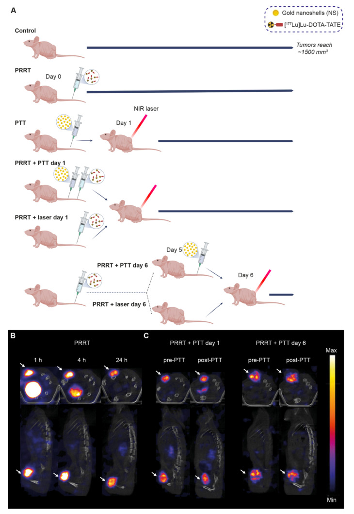
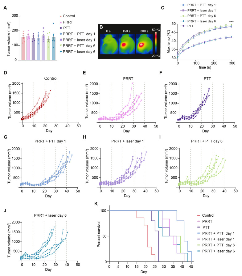
Peptide receptor radionuclide therapy (PRRT) relies on α- and β-emitting radionuclides bound to a peptide that commonly targets somatostatin receptors (SSTRs) for the localized killing of tumors through ionizing radiation. A Lutetium-177 (177Lu)-based probe linked to the somatostatin analog octreotate ([177Lu]Lu-DOTA-TATE) is approved for the treatment of certain SSTR-expressing tumors and has been shown to improve survival. However, a limiting factor of PRRT is the potential toxicity derived from the high doses needed to kill the tumor. This could be circumvented by combining PRRT with other treatments for an enhanced anti-tumor effect. Photothermal therapy (PTT) relies on nanoparticle-induced hyperthermia for cancer treatment and could be a useful add-on to PRRT. Here, we investigate a strategy combining [177Lu]Lu-DOTA-TATE PRRT and nanoshell (NS)-based PTT for the treatment of SSTR-expressing small-cell lung tumors in mice. Our results showed that the combination treatment improved survival compared to PRRT alone, but only when PTT was performed one day after [177Lu]Lu-DOTA-TATE injection (one of the timepoints examined), showcasing the effect of treatment timing in relation to outcome. Furthermore, the combination treatment was well-tolerated in the mice. This indicates that strategies involving NS-based PTT as an add-on to PRRT could be promising and should be investigated further.
Keywords: [177Lu]Lu-DOTA-TATE; cancer; nanoshells (NS); peptide receptor radionuclide therapy (PRRT); photothermal therapy (PTT); somatostatin receptor (SSTR).
Conflict of interest statement
The authors declare no conflict of interest. The company had no role in the design of the study; in the collection, analyses, or interpretation of data; in the writing of the manuscript, and in the decision to publish the results.
Figures


Similar articles
-
Overcoming nephrotoxicity in peptide receptor radionuclide therapy using [177Lu]Lu-DOTA-TATE for the treatment of neuroendocrine tumours.Nucl Med Biol. 2021 Nov-Dec;102-103:1-11. doi: 10.1016/j.nucmedbio.2021.06.006. Epub 2021 Jul 1. Nucl Med Biol. 2021. PMID: 34242948 Review.
-
Evaluation of the Efficacy of a Combined Treatment Using the mTOR-Inhibitor Everolimus and [177Lu]Lu-DOTA-TATE in Nude CD1 Mice with SSTR-Expressing Pancreatic AR42J Xenograft Tumors.Biomedicines. 2022 Dec 1;10(12):3102. doi: 10.3390/biomedicines10123102. Biomedicines. 2022. PMID: 36551858 Free PMC article.
-
Safety, Pharmacokinetics, and Dosimetry of a Long-Acting Radiolabeled Somatostatin Analog 177Lu-DOTA-EB-TATE in Patients with Advanced Metastatic Neuroendocrine Tumors.J Nucl Med. 2018 Nov;59(11):1699-1705. doi: 10.2967/jnumed.118.209841. Epub 2018 Apr 13. J Nucl Med. 2018. PMID: 29653971 Free PMC article. Clinical Trial.
-
Estimation of Absorbed Doses of Indigenously Produced "Direct-route" Lutetium-177-[177Lu]Lu-DOTA-TATE PRRT in Normal Organs and Tumor Lesions in Patients of Metastatic Neuroendocrine Tumors: Comparison with No-Carrier-Added [177Lu]Lu-DOTA-TATE and the Trend with Multiple Cycles.Cancer Biother Radiopharm. 2022 Apr;37(3):214-225. doi: 10.1089/cbr.2021.0340. Epub 2021 Dec 15. Cancer Biother Radiopharm. 2022. PMID: 34910891
-
Lutathera®: The First FDA- and EMA-Approved Radiopharmaceutical for Peptide Receptor Radionuclide Therapy.Pharmaceuticals (Basel). 2019 Jul 29;12(3):114. doi: 10.3390/ph12030114. Pharmaceuticals (Basel). 2019. PMID: 31362406 Free PMC article. Review.
Cited by
-
Effective Treatment of SSTR2-Positive Small Cell Lung Cancer Using 211At-Containing Targeted α-Particle Therapy Agent Which Promotes Endogenous Antitumor Immune Response.Mol Pharm. 2023 Nov 6;20(11):5543-5553. doi: 10.1021/acs.molpharmaceut.3c00427. Epub 2023 Oct 3. Mol Pharm. 2023. PMID: 37788300 Free PMC article.
-
Advances in Radionuclides and Radiolabelled Peptides for Cancer Therapeutics.Pharmaceutics. 2023 Mar 17;15(3):971. doi: 10.3390/pharmaceutics15030971. Pharmaceutics. 2023. PMID: 36986832 Free PMC article. Review.
-
Enhancing [177Lu]Lu-DOTA-TATE therapeutic efficacy in vitro by combining it with metronomic chemotherapeutics.EJNMMI Res. 2024 Aug 13;14(1):73. doi: 10.1186/s13550-024-01135-0. EJNMMI Res. 2024. PMID: 39136880 Free PMC article.
-
Radionuclide-labelled nanoparticles for cancer combination therapy: a review.J Nanobiotechnology. 2024 Nov 22;22(1):728. doi: 10.1186/s12951-024-03020-3. J Nanobiotechnology. 2024. PMID: 39578828 Free PMC article. Review.
References
-
- Bison S.M., Konijnenberg M.W., Melis M., Pool S.E., Bernsen M., Teunissen J.J.M., Kwekkeboom D.J., De Jong M. Peptide receptor radionuclide therapy using radiolabeled somatostatin analogs: Focus on future developments. Clin. Transl. Imaging. 2014;2:55–66. doi: 10.1007/s40336-014-0054-2. - DOI - PMC - PubMed
Grants and funding
LinkOut - more resources
Full Text Sources
Miscellaneous
