. 2022 Jun 16;14(6):1286.
doi: 10.3390/pharmaceutics14061286.

Novel In Situ-Cross-Linked Electrospun Gelatin/Hydroxyapatite Nonwoven Scaffolds Prove Suitable for Periodontal Tissue Engineering

Affiliations

Novel In Situ-Cross-Linked Electrospun Gelatin/Hydroxyapatite Nonwoven Scaffolds Prove Suitable for Periodontal Tissue Engineering

Martin Philipp Dieterle et al. Pharmaceutics. .

Abstract

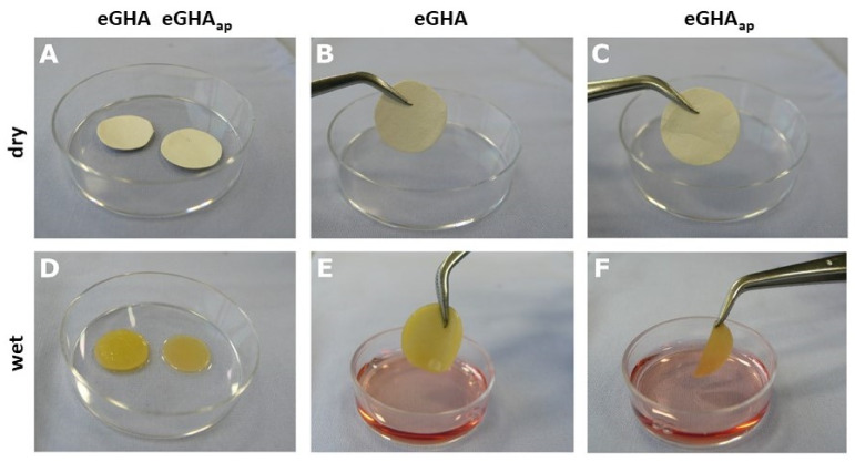
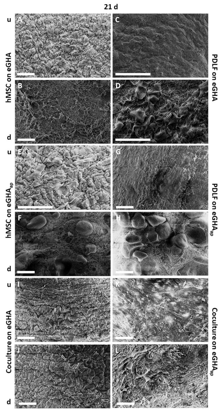
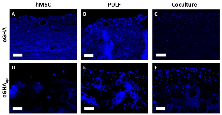
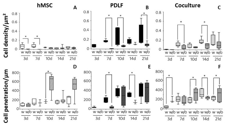
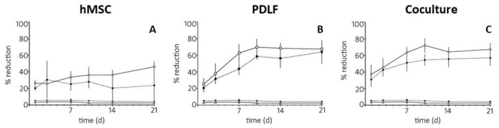
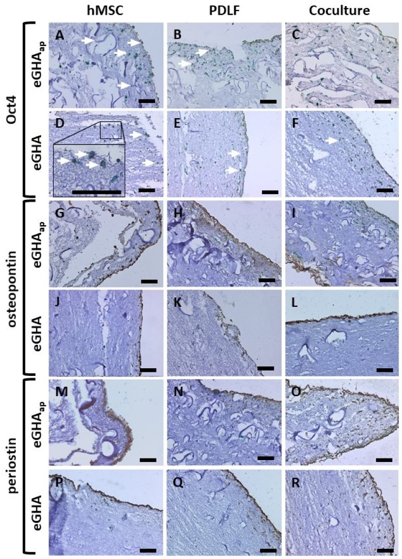
Periodontal diseases affect millions of people worldwide and can result in tooth loss. Regenerative treatment options for clinical use are thus needed. We aimed at developing new nonwoven-based scaffolds for periodontal tissue engineering. Nonwovens of 16% gelatin/5% hydroxyapatite were produced by electrospinning and in situ glyoxal cross-linking. In a subset of scaffolds, additional porosity was incorporated via extractable polyethylene glycol fibers. Cell colonization and penetration by human mesenchymal stem cells (hMSCs), periodontal ligament fibroblasts (PDLFs), or cocultures of both were visualized by scanning electron microscopy and 4',6-diamidin-2-phenylindole (DAPI) staining. Metabolic activity was assessed via Alamar Blue® staining. Cell type and differentiation were analyzed by immunocytochemical staining of Oct4, osteopontin, and periostin. The electrospun nonwovens were efficiently populated by both hMSCs and PDLFs, while scaffolds with additional porosity harbored significantly more cells. The metabolic activity was higher for cocultures of hMSCs and PDLFs, or for PDLF-seeded scaffolds. Periostin and osteopontin expression was more pronounced in cocultures of hMSCs and PDLFs, whereas Oct4 staining was limited to hMSCs. These novel in situ-cross-linked electrospun nonwoven scaffolds allow for efficient adhesion and survival of hMSCs and PDLFs. Coordinated expression of differentiation markers was observed, which rendered this platform an interesting candidate for periodontal tissue engineering.

Keywords: biocompatible materials; gelatin; hydroxyapatites; mesenchymal stem cells; periodontal guided tissue regeneration; periodontal ligament; periodontitis; regenerative medicine; tissue engineering.


Conflict of interest statement

The authors declare no conflict of interest.

Figures

Figure 1
Figure 1
Photographs of electrospun gelatin/hydroxyapatite scaffolds with (eGHAap) and without additional porosity (eGHA). The scaffolds were punched in circles after drying (AC) and subsequently rewetted with culture medium (DF). (AC) If dry, there was no remarkable macroscopic difference between eGHA and eGHAap (A, left: eGHA, right: eGHAap) apart from the slightly papery appearance of eGHAap (C) when compared with that of eGHA (B). (DE) Following wetting and moisture expansion, eGHA appeared clearly thicker (D, left) and inherently more stable (E), while eGHAap collapsed if taken with forceps (F) but remained in shape and easily unfolded if laid down (D, right).
Figure 2
Figure 2
Scanning electron micrographs of eGHA/eGHAap scaffolds after 21 d. Illustrated is always the surface of the scaffolds, indicating the different morphologies of the respective cell types under study which from time to time reveal the nanofibers on the underside of the scaffolds (B,D,F,H,J,L). hMSCs (A,B,E,F), PDLFs (C,D,G,H), or cocultures of both (IL) were seeded on either eGHA (AD,I,J) or eGHAap scaffolds (EH,K,L) and prepared for scanning electron microscopy (SEM) analysis after 21 d. The top sides (u) of the monocultures (A,C,E,G) were densely populated with either hMSCs or PDLFs, while the bottom sides (d) (B,D,F,H) illustrated the geometric configurations of the nonwovens and were barely populated by cells, as expected. In the cocultures, (u) were populated by hMSCs (I,K) and (d) with PDLFs (J,L). Details are given in the main text. All scaffolds, irrespective of the presence of additional porosity, were densely covered with the indicated cells, proving the overall suitability of the eGHA/eGHAap nonwovens for the adhesion and spreading of periodontal fibroblasts and mesenchymal stem cells. The cell morphologies could be described as polygonal or spindle-like. Scale bars represent 100 µm.
Figure 3
Figure 3
Representative cutouts of 4′,6-diamidin-2-phenylindole (DAPI) stained sections from eGHA and eGHAap nonwovens populated with hMSCs, PDLFs, or cocultures after an incubation period of 21 d. The upsides of the monoculture scaffolds are oriented towards the top of the picture. For the cocultures (C,F), the margins of the PDLF-populated downsides are shown. (A) hMSCs, (B) PDLFs, or (C) cocultures were grown on eGHA scaffolds. Cell nuclei are presented as blue dots, while parts of the nonwovens also show some background fluorescence. Accordingly, eGHAap samples populated with (D) hMSCs, (E) PDLFs, or (F) cocultures are presented, showing a tendency towards increased cell densities when compared to eGHA nonwovens. Scale bars represent 100 µm.
Figure 4
Figure 4
Statistical evaluation of the 4′,6-diamidin-2-phenylindole (DAPI) stained sections after incubation for 3, 7, 10, 14, and 21 d. (AC) Quantitative analysis of cell densities (cells/µm2) on nonwovens with (=eGHAap = w) or without (=eGHA = w/o) additional porosity populated by (A) hMSCs, (B) PDLFs, and (C) cocultures of both cell types. The boxplots represent the median values and interquartile ranges. The whiskers depict the 1.5-fold interquartile ranges. PDLFs on d 3 on eGHA could not be evaluated for technical reasons. (D,E) Quantitative analysis of the maximum cell penetration (µm) on nonwovens with (=eGHAap = w) or without (=eGHA = w/o) additional porosity populated by (D) hMSCs, (E) PDLFs, or (F) cocultures of both cell types. The boxplots represent the median values and interquartile ranges. The whiskers depict the 1.5-fold interquartile ranges. Numerical data for the graphs can be found in the Supplementary Tables S1 and S2. *: p < 0.05.
Figure 5
Figure 5
Line graphs representing the metabolic activities of hMSCs, PDLFs, or cocultures of both cell types on eGHA or eGHAap. The resazurin/Alamar Blue® activity assay was used to determine the reductive activity of the cells, which is an indirect measure for cell viability and metabolism. The indicated cells were incubated on the scaffolds for 1, 3, 7, 10, 14, and 21 d. Completely reduced Alamar Blue® reagent was used as a positive control (=100%). eGHA (black triangles) and eGHAap (white triangles) without cells were used as negative controls for all experimental conditions. Mean metabolic activities and the corresponding standard deviations (SD) are depicted. The numerical data are presented in Supplementary Table S3. (A) Comparison of the reductive capacity of hMSCs grown on eGHA (black rhombs) and eGHAap (white rhombs). (B) Comparison of the reductive capacity of PDLFs grown on on eGHA (black squares) and eGHAap (white squares). (C) Comparison of the reductive capacity of interactive cocultures grown on eGHA (black circles) and eGHAap (white circles).
Figure 6
Figure 6
Immunohistochemical evaluation of eGHA and eGHAap scaffolds populated with hMSCs, PDLFs, and cocultures of both cell types. After an incubation period of 21 d, the constructs were fixed and incubated with primary antibodies against Oct4 (AF), osteopontin (GL), and periostin (MR) and stained with the peroxidase-dependent 3,5-diaminobenzidine reaction (DAB; brown color). Subsequently, immunohistochemical staining for the mesenchymal cytoskeletal filament vimentin (HistoGreen staining; green color) was performed for all sections to detect all cells. Finally, hematoxylin counterstaining was applied. For each protein of interest, hMSCs (A,D,G,J,M,P), PDLFs (B,E,H,K,N,Q), and cocultures (C,F,I,L,O,R) grown on either eGHAap (AC,GI,MO) or eGHA (DF,JL,PR) were examined. The orientation of the representative pictures follows the same rules as in Figure 3. Staining results were evaluated visually and only qualitatively. (A,D) Oct4 was expressed mainly in hMSCs and detectable in only a few PDLFs (B,E). The inset in (D) represents a higher magnification of the section, illustrating the nuclear localization of Oct4. Osteopontin, a marker protein of hard tissues, was expressed predominantly in the nonwovens populated with cocultures (I,L). The periodontium-related marker periostin was found mainly in the cocultures (O,R) or PDLFs (N,Q), but barely in hMSCs (M,P). The white arrows in (A,B,D,F) exemplarily indicate positively stained cells, which are otherwise hard to recognize. Scale bars represent 100 µm.

Similar articles

Cited by

References

    1. Fan C., Li Z., Ji Q., Sun H., Liang Y., Yang P. Carboxymethyl chitin or chitosan for osteoinduction effect on the human periodontal ligament stem cells. Dent. Mater. J. 2022;41:392–401. doi: 10.4012/dmj.2021-250. - DOI - PubMed
    1. Guo Y., Wang X., Wang C., Chen S. In vitro behaviour of human gingival fibroblasts cultured on 3d-printed titanium alloy with hydrogenated tio2 nanotubes. J. Mater. Sci. Mater. Med. 2022;33:1–11. doi: 10.1007/s10856-022-06649-4. - DOI - PMC - PubMed
    1. Komatsu K., Ideno H., Shibata T., Nakashima K., Nifuji A. Platelet-derived growth factor-bb regenerates functional periodontal ligament in the tooth replantation. Sci. Rep. 2022;12:1–16. doi: 10.1038/s41598-022-06865-6. - DOI - PMC - PubMed
    1. Rosin F.C.P., Gonsalves H., Santos A.F., de Paula Novaes C., Huang I., Deboni M.C.Z., Corrêa L. Keratin expression in gingival tissue and primary cultured gingival keratinocytes: Are there differences? Arch. Oral Biol. 2020;117:104780. doi: 10.1016/j.archoralbio.2020.104780. - DOI - PubMed
    1. Dieterle M.P., Husari A., Steinberg T., Wang X., Ramminger I., Tomakidi P. From the matrix to the nucleus and back: Mechanobiology in the light of health, pathologies, and regeneration of oral periodontal tissues. Biomolecules. 2021;11:824. doi: 10.3390/biom11060824. - DOI - PMC - PubMed