. 2022 May 30;10(6):875.
doi: 10.3390/vaccines10060875.

Epitope-Based Vaccines against the Chlamydia trachomatis Major Outer Membrane Protein Variable Domain 4 Elicit Protection in Mice

Affiliations

Epitope-Based Vaccines against the Chlamydia trachomatis Major Outer Membrane Protein Variable Domain 4 Elicit Protection in Mice

Amanda L Collar et al. Vaccines (Basel). .

Abstract

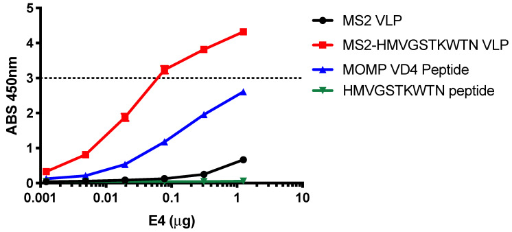
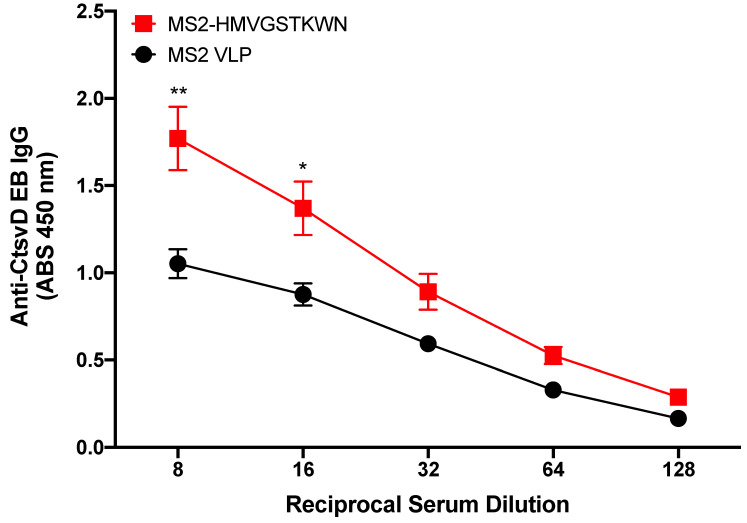
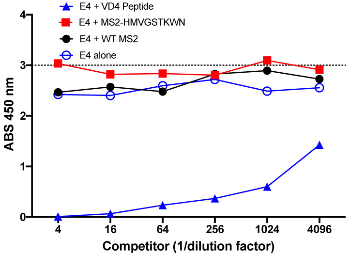
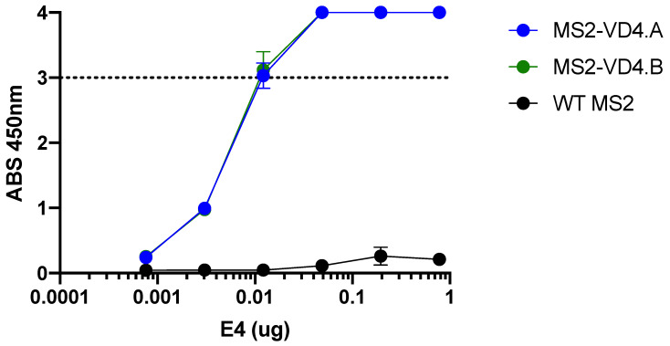
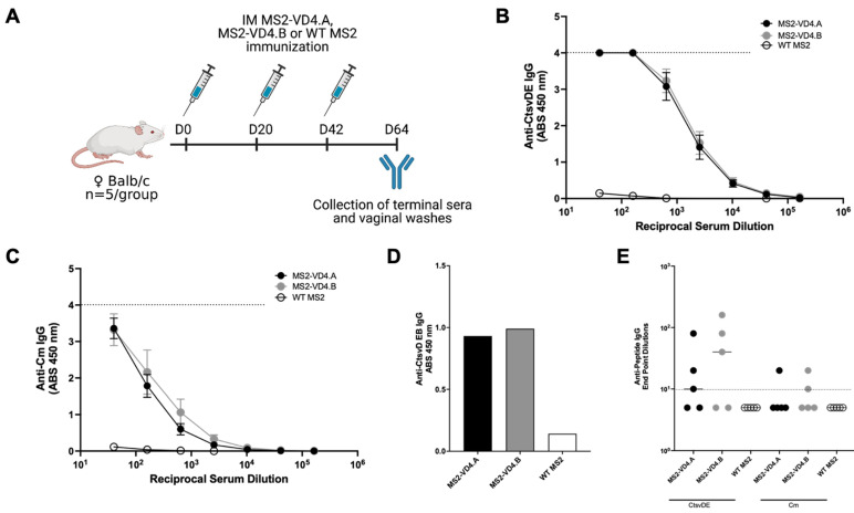
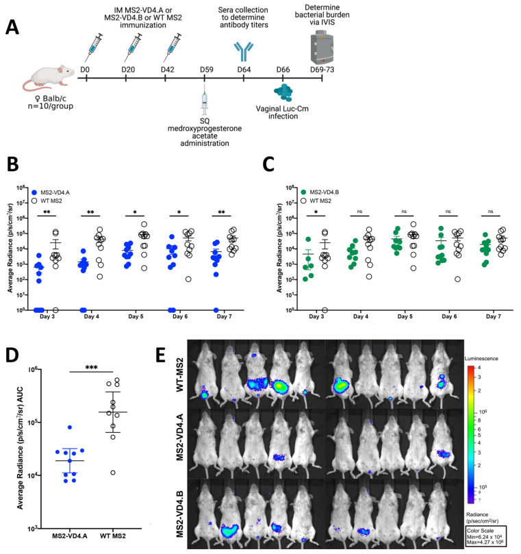
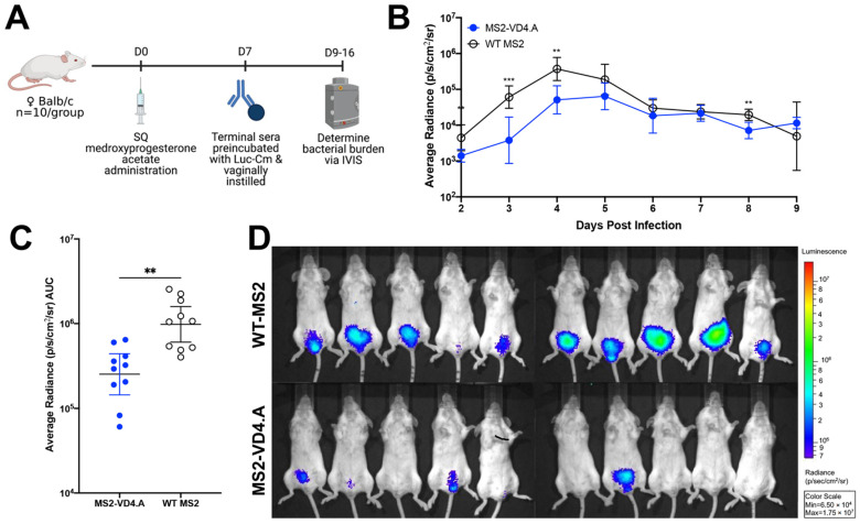
Chlamydia trachomatis (Ct) is the most common bacterial sexual transmitted pathogen, yet a vaccine is not currently available. Here, we used the immunogenic bacteriophage MS2 virus-like particle (VLP) technology to engineer vaccines against the Ct major outer membrane protein variable domain 4 (MOMP-VD4), which contains a conserved neutralizing epitope (TTLNPTIAG). A previously described monoclonal antibody to the MOMP-VD4 (E4 mAb) is capable of neutralizing all urogenital Ct serovars and binds this core epitope, as well as several non-contiguous amino acids. This suggests that this core epitope may require conformational context in order to elicit neutralizing antibodies to Ct. In order to identify immunogens that could elicit neutralizing antibodies to the TTLNPTIAG epitope, we used two approaches. First, we used affinity selection with a bacteriophage MS2-VLP library displaying random peptides in a constrained, surface-exposed loop to identify potential E4 mAb mimotopes. After four rounds of affinity selection, we identified a VLP-displayed peptide (HMVGSTKWTN) that could bind to the E4 mAb and elicited serum IgG that bound weakly to Ct elementary bodies by ELISA. Second, two versions of the core conserved TTLNPTIAG epitope (TTLNPTIAG and TTLNPTIAGA) were recombinantly expressed on the coat protein of the MS2 VLP in a constrained, surface-exposed loop. Mouse immune sera IgG bound to Ct elementary bodies by ELISA. Immunization with these MS2 VLPs provided protection from vaginal Chlamydia infection in a murine challenge model. These data suggest that short peptide epitopes targeting the MOMP-VD4 could be appropriate for Ct vaccine design when displayed on an immunogenic bacteriophage VLP vaccine platform.

Keywords: Chlamydia trachomatis; affinity selection; antibodies; bacteriophage; epitope; vaccine; virus-like particle.


Conflict of interest statement

A.L.C. and K.M.F. are inventors on a patent pending for vaccines described in this manuscript. The funders had no role in the design of the study; in the collection, analyses, or interpretation of data; in the writing of the manuscript, or in the decision to publish the results.

Figures

Figure 1
Figure 1
Schematic of MS2 bacteriophage VLP-based approaches for MOMP VD4 conserved epitope vaccine design. (A) MS2 bacteriophage coat protein dimers each display one foreign peptide in the surface-exposed beta-hairpin, called the AB loop. (B) The sequence of the VD4 core conserved epitope (TTLNPTIAG) is shown in alignment for all urogenital Ct serovars (* perfect conservation), as well as Cm. Cm alignment scores are as compared to CtsvD (shown below Cm sequence), and alignment scores of all urogenital serovars are shown below Ct SvK. (C) Affinity selection approach against E4 mAb. An MS2 VLP library displaying random peptides in the AB loop is panned against the E4 mAb. Bound VLPs are eluted, and encapsidated coding RNA is extracted. This is then subjected to RT-PCR and cloning into the MS2 VLP expression plasmid, which is then transformed into E. coli to produce a new VLP library that is enriched for VLPs that bind to E4 mAb. The process is then repeated for four total rounds. The RT-PCR product from the final round is then subjected to deep sequencing to identify peptides of interest that bind to E4 mAb. (D) Rational-design approach using display of the VD4 core conserved epitope MS2 VLPs. The coding sequences for peptides are cloned into an MS2 coat protein expression plasmid such that the peptide is displayed in the AB loop. Plasmids are then transformed into E. coli, and VLPs are expressed and purified.
Figure 2
Figure 2
E4 mAb binds to HMVGSTKWTN peptide in a conformation-dependent manner. E4 mAb binding to linear HMVGSTKWTN peptide, MS2-HMVGSTKWTN (MS2 VLP displaying HMVGSTKWTN in the AB loop), MS2 VLP (negative control), and VD4 epitope linear peptide (FDTTTLNPTIAGAGDVK) was assessed by ELISA at various concentrations of E4 mAb. Data are the average of three technical replicates. The dashed line represents the linear limit of the ELISA plate reader.
Figure 3
Figure 3
Binding characteristics of MS2-HMVGSTKWTN immunized mouse sera IgG to Ct serovar D. Mice (n = 5) were immunized with MS2-HMVGSTKWTN or WT MS2 (negative control). Sera were collected, and binding activity was assessed against Ct serovar D by EB ELISA. Statistical analysis was performed utilizing a nonparametric Mann–Whitney t-test. Quantitative data represent the mean ± SEM. * p < 0.05, ** p < 0.01.
Figure 4
Figure 4
Preferential binding of E4 mAb. Competition peptide ELISA revealed that addition of MS2-HMVGSTKWN as a binding competitor of E4 mAb did not change the binding potential to the VD4 peptide, whereas addition of the linear VD4 peptide reduced binding of the E4 mAb to the bait VD4 peptide.
Figure 5
Figure 5
E4 mAb binds to MS2 VLPs displaying the VD4 epitope. E4 mAb binding capacity to MS2-VD4.A and MS2-VD4.B as measured by ELISA, demonstrating binding above that of WT MS2 across a range of dilutions. Quantitative data represent the mean ± SD.
Figure 6
Figure 6
MS2-VD4 immunization results in high-titer, epitope-specific IgG antibody responses. (A) Immunization schedule. Female Balb/c mice (n = 5/group) were immunized three times, 3 weeks apart with their respective vaccine before collection of terminal immune sera and vaginal washes to investigate antibody responses via ELISA. (B) Binding capacity of MS2-VD4 immune sera IgG to the CtsvDE VD4 epitope peptide (FDTTTLNPTIAGAGDVK). (C) Binding capacity of MS2-VD4 immune sera IgG to the Cm VD4 epitope peptide (LDVTTWNPTIAGAGTIA). (D) Binding capacity of MS2-VD4 immune sera IgG to CtsvD EBs by ELISA. Serum dilution 1:128. (E) Binding capacity of mucosal IgG from vaginal washes to CtsvDE (left) and Cm (right) VD4 epitope peptide. Quantitative data represent the mean ± SEM (B,C) and median (E).
Figure 7
Figure 7
Immunization with MS2-VD4.A results in decreased urogenital Chlamydia burden as compared to WT MS2 immunized controls. (A) Immunization schedule. Female Balb/c mice (n = 10/group) were immunized three times, 3 weeks apart with their respective vaccine. Mice were administered 2.5 mg of Depo-Provera before vaginal challenge with Luc-Cm. (B) Average radiance (p/s/cm2/sr) measured for MS2-VD4.A and WT MS2 on days 3 through 7 post infection. (C) Average radiance (p/s/cm2/sr) measured for MS2-VD4.B and WT MS2 on days 3 through 7 post infection. (D) Area under the curve (AUC) measured for average radiance on days 3 through 7 post infection for MS2-VD4.A and WT MS2. (E) IVIS images for WT MS2 control mice (top), MS2-VD4.A mice (middle), and MS2-VD4.B mice (bottom) on day 7 post infection, with luminescence visualized, shown as a representative image of those collected each day. Statistical analysis was performed utilizing the nonparametric Mann–Whitney t-test. Quantitative data represent the geometric mean ± SEM. * p < 0.05, ** p < 0.01, *** p < 0.001.
Figure 8
Figure 8
Protection from vaginal Chlamydia infection can be mediated by antibodies. (A) Study design for in vivo neutralization. Naïve female Balb/c mice (n = 10/group) were vaginally challenged with 2 × 10⁴ IFU of Luc-Cm that was preincubated with terminal immune sera from mice vaccinated with either MS2-VD4.A or WT MS2. Bacterial burden was determined on days 2 through 9 post infection. (B) Average radiance (p/s/cm2/sr) measured for mice receiving Luc-Cm preincubated with either MS2-VD4.A or WT MS2 immune sera on days 2 through 9 post infection. (C) Area under the curve (AUC) measured for average radiance on days 2 through 9 post infection. (D) IVIS image for mice receiving Luc-Cm preincubated with either WT MS2 immune sera (top) or MS2-VD4.A immune sera (bottom) on day 3 post infection, with luminescence visualized, shown as a representative image of those collected each day. Statistical analysis was performed utilizing the nonparametric Mann–Whitney t-test. Quantitative data represent the geometric mean ± SEM. ** p < 0.01, *** p < 0.001.

Similar articles

Cited by

References

    1. Gottlieb S.L., Deal C.D., Giersing B., Rees H., Bolan G., Johnston C., Timms P., Gray-Owen S.D., Jerse A.E., Cameron C.E., et al. The Global Roadmap for Advancing Development of Vaccines against Sexually Transmitted Infections: Update and next Steps. Vaccine. 2016;34:2939–2947. doi: 10.1016/j.vaccine.2016.03.111. - DOI - PMC - PubMed
    1. Broutet N., Fruth U., Deal C., Gottlieb S.L., Rees H. Vaccines against Sexually Transmitted Infections: The Way Forward. Vaccine. 2014;32:1630–1637. doi: 10.1016/j.vaccine.2014.01.053. - DOI - PubMed
    1. Zhong G., Brunham R.C., de la Maza L.M., Darville T., Deal C. National Institute of Allergy and Infectious Diseases Workshop Report: “Chlamydia Vaccines: The Way Forward”. Vaccine. 2019;37:7346–7354. doi: 10.1016/j.vaccine.2017.10.075. - DOI - PubMed
    1. Caldwell H.D., Schachter J. Antigenic Analysis of the Major Outer Membrane Protein of Chlamydia spp. Infect. Immun. 1982;35:1024–1031. doi: 10.1128/iai.35.3.1024-1031.1982. - DOI - PMC - PubMed
    1. Yuan Y., Zhang Y.X., Watkins N.G., Caldwell H.D. Nucleotide and Deduced Amino Acid Sequences for the Four Variable Domains of the Major Outer Membrane Proteins of the 15 Chlamydia trachomatis Serovars. Infect. Immun. 1989;57:1040–1049. doi: 10.1128/iai.57.4.1040-1049.1989. - DOI - PMC - PubMed

LinkOut - more resources