. 2022 Jun 10;10(6):929.
doi: 10.3390/vaccines10060929.

Immunological Study of Combined Administration of SARS-CoV-2 DNA Vaccine and Inactivated Vaccine

Affiliations

Immunological Study of Combined Administration of SARS-CoV-2 DNA Vaccine and Inactivated Vaccine

Ziyan Meng et al. Vaccines (Basel). .

Abstract

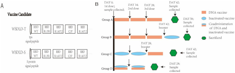
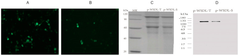
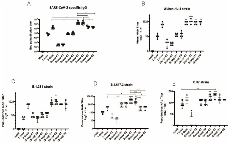
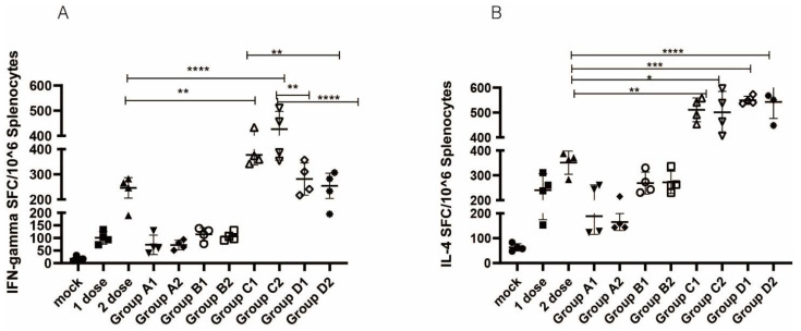
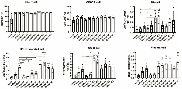
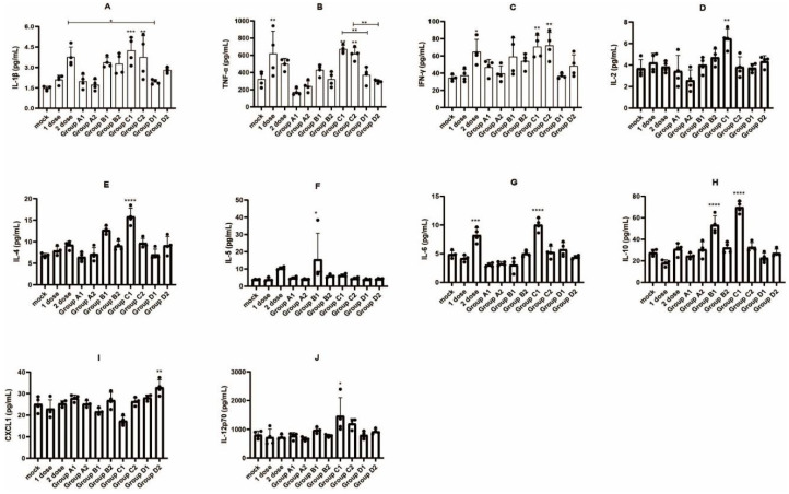
Objective: We constructed two DNA vaccines containing the receptor-binding domain (RBD) genes of multiple SARS-CoV-2 variants and used them in combination with inactivated vaccines in a variety of different protocols to explore potential novel immunization strategies against SARS-CoV-2 variants. Methods: Two DNA vaccine candidates with different signal peptides (namely, secreted and membrane signal peptides) and RBD protein genes of different SARS-CoV-2 strains (Wuhan-Hu-1, B.1.351, B.1.617.2, C.37) were used. Four different combinations of DNA and inactivated vaccines were tested, namely, Group A: three doses of DNA vaccine; B: three doses of DNA vaccine and one dose of inactivated vaccine; C: two doses of inactivated vaccine and one dose of DNA vaccine; and D: coadministration of DNA and inactivated vaccines in two doses. Subgroups were grouped according to the signal peptide used (subgroup 1 contained secreted signal peptides, and subgroup 2 contained membrane signal peptides). The in vitro expression of the DNA vaccines, the humoral and cellular immunity responses of the immunized mice, the immune cell population changes in local lymph nodes, and proinflammatory cytokine levels in serum samples were evaluated. Results: The antibody responses and cellular immunity in Group A were weak for all SARS-CoV-2 strains; for Group B, there was a great enhancement of neutralizing antibody (Nab) titers against the B.1.617.2 variant strain. Group C showed a significant increase in antibody responses (NAb titers against the Wuhan-Hu-1 strain were 768 and 1154 for Group C1 and Group C2, respectively, versus 576) and cellular immune responses, especially for variant B.1.617.2 (3240 (p < 0.001) and 2430 (p < 0.05) for Group C1 and Group C2, versus 450); Group D showed an improvement in immunogenicity. Group C induced higher levels of multiple cytokines. Conclusion: The DNA vaccine candidates we constructed, administered as boosters, could enhance the humoral and cellular immune responses of inactivated vaccines against COVID-19, especially for B.1.617.2.

Keywords: COVID-19; DNA vaccine; RBD; SARS-CoV-2; inactivated vaccine; variants.


Conflict of interest statement

The authors declare that they have no competing financial interests or personal relationships that could have appeared to influence the work reported in this paper.

Figures

Figure 1
Figure 1
(A) Schematic diagram of two DNA vaccine candidate constructs. RBD: receptor binding domain. (B) Animal immunization and sample collection procedures. The DNA vaccines all contained two RBD genes of B.1.617.2 (Delta strain).
Figure 2
Figure 2
Protein expression analysis: (A) The expression of recombinant plasmid WSDL-T-EGFP in transfected HEK293T cells. (B) The expression of recombinant plasmid WSDL-S-EGFP in transfected HEK293T cells. Images were analyzed with a fluorescence microscope with a 20× objective. (C) SDS–PAGE and (D) western blot assay to detect the purified protein of DNA vaccines (p-WSDL-T and p-WSDL-S) bearing four RBD regions expressed in ExpiCHO cell lines.
Figure 3
Figure 3
Antibody responses to different group designs: (A) Titers of SARS-SoV-2 S1-specific total IgG analysis (Wuhan-Hu-1 strain). (BE) Neutralizing antibody (NAb) titers against the (B) Wuhan-Hu-1, (C) B.1.351, (D) B.1.617.2, and (E) C.37 strains. The latter three variants were tested by pseudovirus neutralization assays. 1 dose: 1 dose inactivated vaccine control group; 2 dose: 2 doses inactivated vaccine control group; one-way ANOVA (and nonparametric or mixed) was conducted; * p < 0.05; ** p < 0.01; *** p < 0.001; **** p < 0.0001 versus the control group.
Figure 4
Figure 4
SARS-CoV-2 RBD-specific T-cell responses analyzed by ELISPOT: (A) RBD-specific IFN-γ and (B) RBD-specific IL-4 responses were measured. 1 dose: 1 dose inactivated vaccine control group; 2 dose: 2 doses inactivated vaccine control group; one-way ANOVA (and nonparametric or mixed) was conducted; * p < 0.05; ** p < 0.01; *** p < 0.001; **** p < 0.0001 versus the control group.
Figure 5
Figure 5
Different vaccination groups elicited immune cell population percentage changes in CD4+ T cells, CD8+ T cells, Tfh cells, GC B cells, plasma cells and IFN-γ-secreting cells in local lymph nodes. For cell data detected as 0% in flow cytometry, we assigned a value of 0.05% for plotting. 1 dose: 1 dose inactivated vaccine control group; 2 dose: 2 doses inactivated vaccine control group; one-way ANOVA (and nonparametric or mixed) was conducted; * p < 0.05; ** p < 0.01; *** p < 0.001; **** p < 0.0001 versus the control group.
Figure 6
Figure 6
Proinflammatory cytokine analysis of serum samples collected from mice at the time of sacrificing after completion of the immunization procedure. (A) IL-1β, (B) TNF-α, (C) IFN-γ, (D) IL-2, (E) IL-4, (F) IL-5, (G) IL-6, (H) IL-10, (I) CXCL1, and (J) IL-12p70. 1 dose: 1 dose inactivated vaccine control group; 2 dose: 2 doses inactivated vaccine control group; one-way ANOVA (and nonparametric or mixed) was conducted; * p < 0.05; ** p < 0.01; *** p < 0.001; **** p < 0.0001 versus the control group.

Similar articles

Cited by

References

    1. Mackenzie J.S., Smith D.W. COVID-19: A novel zoonotic disease caused by a coronavirus from China: What we know and what we don’t. Microbiol. Aust. 2020;41:45. doi: 10.1071/MA20013. - DOI - PMC - PubMed
    1. World Health Organization Virtual Press Conference on COVID-19. 2020. [(accessed on 21 April 2022)]. Available online: https://www.who.int/docs/default-source/coronaviruse/transcripts/who-aud....
    1. World Health Organization Home/Diseases/Coronavirus Disease (COVID-19) [(accessed on 21 April 2022)]. Available online: https://www.who.int/emergencies/diseases/novel-coronavirus-2019.
    1. Lu R., Zhao X., Li J., Niu P., Yang B., Wu H., Wang W., Song H., Huang B., Zhu N., et al. Genomic characterisation and epidemiology of 2019 novel coronavirus: Implications for virus origins and receptor binding. Lancet. 2020;395:565–574. doi: 10.1016/S0140-6736(20)30251-8. - DOI - PMC - PubMed
    1. Kim D., Lee J.Y., Yang J.S., Kim J.W., Kim V.N., Chang H. The Architecture of SARS-CoV-2 Transcriptome. Cell. 2020;181:914–921. doi: 10.1016/j.cell.2020.04.011. - DOI - PMC - PubMed

LinkOut - more resources