. 2022 Jun 17;10(6):969.
doi: 10.3390/vaccines10060969.

Early IgE Production Is Linked with Extrafollicular B- and T-Cell Activation in Low-Dose Allergy Model

Affiliations

Early IgE Production Is Linked with Extrafollicular B- and T-Cell Activation in Low-Dose Allergy Model

Dmitrii Borisovich Chudakov et al. Vaccines (Basel). .

Abstract

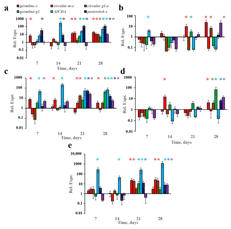
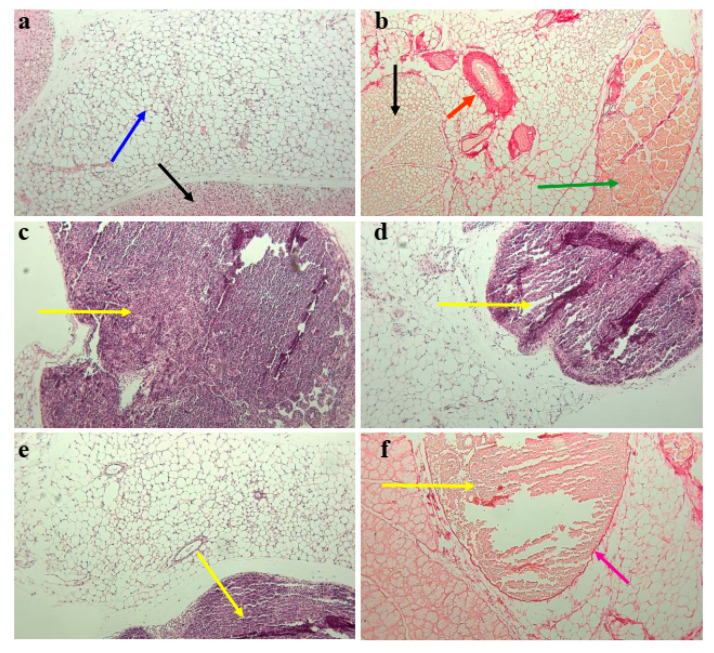
Despite its paramount importance, the predominant association of early IgE production with harmless antigens, via germinal-center B- and T-cell subpopulations or extrafollicular activation, remains unresolved. The aim of this work was to clarify whether the reinforced IgE production following the subcutaneous immunization of BALB/c mice with low antigen doses in withers adipose tissue might be linked with intensified extrafollicular or germinal-center responses. The mice were immunized three times a week for 4 weeks in the withers region, which is enriched in subcutaneous fat and tissue-associated B cells, with high and low OVA doses and via the intraperitoneal route for comparison. During long-term immunization with both low and high antigen doses in the withers region, but not via the intraperitoneal route, we observed a significant accumulation of B220-CD1d-CD5-CD19+ B-2 extrafollicular plasmablasts in the subcutaneous fat and regional lymph nodes but not in the intraperitoneal fat. Only low antigen doses induced a significant accumulation of CXCR4+ CXCR5- CD4+ extrafollicular T helpers in the withers adipose tissue but not in the regional lymph nodes or abdominal fat. Only in subcutaneous fat was there a combination of extrafollicular helper accumulation. In conclusion, extrafollicular B- and T-cell activation are necessary for early IgE class switching.

Keywords: IgE; extrafollicular T helpers; extrafollicular response; plasmablasts; subcutaneous fat.


Conflict of interest statement

The authors declare no conflict of interest.

Figures

Figure 1
Figure 1
Specific IgE production was induced rapidly after immunization by the SC route in the withers region but not after IP immunization. It was accompanied by relatively low IgG1 production and correlated with systemic anaphylaxis after the administration of low antigen doses. BALB/c mice (n = 4 for each group–time point) were immunized by the SC route in the withers region (ac), (g,h) or by the IP route (df), with indicated OVA doses for 4 weeks 3 times a week. Specific IgE (a,d) and IgG1 (b,e) production was measured at the indicated time points, as was the systemic anaphylaxis intensity 45 min after the administration of the resolving antigen dose (250 µg) (c,f). The correlation of the IgE titers with systemic anaphylaxis (g,h). Blue asterisks signify the differences between low-dose-SC-immunized mice and the intact group (*/** signify p values < 0.05/0.01 correspondingly); red ones signify the differences between high-dose-SC-immunized mice and the intact group; green ones signify the differences between low-dose-IP-immunized mice and the intact group. All the experiments were performed 3 times, and representative data are shown.
Figure 2
Figure 2
Antigen administration induced Ig class switching more rapidly in withers fat tissue compared to lymph nodes or abdominal fat. BALB/c mice (n = 4 for each group–time point) were immunized by the SC route in the withers region (ad) or by the IP route (e) with the indicated OVA doses for 4 weeks, 3 times a week. The expression of the indicated genes and transcripts in the indicated time points are shown for low- (a,b,e) and high-antigen-dose (c,d)-immunized mice in withers subcutaneous fat (a,c), regional lymph nodes close to the withers region (b,d), and abdominal fat (e). Each asterisk indicates significant differences (p < 0.05) in gene expression shown by the same color with an intact group. Each asterisk of a certain color corresponds to the significance of the difference of the gene expression, which is shown by a column of the same color. All experiments were performed 3 times, and representative data are shown.
Figure 3
Figure 3
Subcutaneous fat adipose tissue in the withers region contained large bordered tertiary lymphoid structures. Representative histological images of mouse subcutaneous withers adipose tissue. Tissue samples were taken from low-dose-immunized mice 4 weeks after the start of antigen administration. Samples were stained with H and E (a,ce) or Picro Sirius Red (b,f). Adipose tissue in intact mice was of two types: white, as indicated by the blue arrow in (a), and brown, as indicated by the black arrow in (a). The Picro Sirius Red-stained vessel is indicated by the red arrow in (b); the muscles, by the green arrow in (b); and the brown adipose cells, by the black arrow in (b). Large clusters of lymphoid cells are indicated by the yellow arrows in (cf), stained with H and E (C–E) or Picro Sirius Red (f). The fibrotic border of the cluster is indicated by the purple arrow in (f). Magnification: 100×.
Figure 4
Figure 4
Low antigen doses triggered B220− CD38−/+ CD95+ extrafollicular plasmablasts, but not classical germinal center accumulation in withers adipose tissue and lymph nodes. BALB/c mice (n = 4 for each group–time point) were immunized by the SC route in the withers region (ag) or by the IP route (e) with low (100 ng) or high (10,000 ng) OVA doses for 4 weeks, 3 times a week. Representative flow cytometry pseudocolor plots from mouse subcutaneous fat tissue cells gated on CD19+ Cd5- CD1d- B-2 B cells (a), B220− B-2 plasmablasts (b), and B220+ non-plasmablast B-2 B cells (c). Roman numerals correspond to the following subpopulations: I—B220- plasmablasts; II—B220+ non-plasmablast B-2 B cells; II–V—different plasmablast subpopulations; VI—CD38- CD95+ classical germinal centers; VII—CD38+ CD95+ activated B-2 B cells. The percentage of B220+ CD38− CD95+ classical germinal centers and B220− CD38− CD95+ and B220− CD38+ CD95+ plasmablasts at the indicated time points are shown for low (d,e,h)- and high-antigen dose (e,g)-immunized mice in withers subcutaneous fat (d,f), regional lymph nodes close to the withers region (e,g), and abdominal fat (e). Each asterisk indicates significant differences (p < 0.05) in the percentages of the subpopulation in B cells shown by the same color with the intact group. Each asterisk of a certain color corresponds to the significance of the difference of subpopulation percentage, which is shown by a column of the same color. All experiments were performed 3 times, and representative data are shown.
Figure 5
Figure 5
Proportions of extrafollicular plasmablast subpopulations correlated with IgE production. BALB/c mice (n = 4 for each group–time point) were immunized by the SC route in the withers region with the indicated OVA doses for 4 weeks, 3 times a week. Correlation of the percentage of CD38− CD95+ (a,d) and CD38+ CD95+ (b,e) extrafollicular plasmablasts in B cells and B220+ CD38− CD95+ classical germinal centers (c,f), in withers adipose fat (ac), and regional lymph nodes (df). All the experiments were performed 3 times, and representative data are shown.
Figure 6
Figure 6
Unique pattern of humoral immune response and early induction of IgE B-cell class switching in withers subcutaneous fat upon low-antigen-dose administration may be linked with accumulation of extrafollicular T helpers and the absence of the accumulation of T helpers associated with B-cell follicles and follicular T helpers. BALB/c mice (n = 4 for each group–time point) were immunized by the SC route in the withers region (ae) or by the IP route (f) with low (100 ng) or high (10,000 ng) OVA doses for 4 weeks, 3 times a week. Representative flow cytometry pseudocolor plots of subcutaneous fat cells gated on CD4+ CD49b- CD45+ T helpers (a). Percentage of CD4+ CXCR4-CXCR5+ T helpers situated in B-cell follicles, CD4+ CXCR4+ CXCR5+ follicular T helpers, and CD4+ CXCR4+ CXCR5- extrafollicular T helpers at indicated time points are shown for low (b,c,f)- and high-antigen-dose (d,e)-immunized mice in withers subcutaneous fat (b,d), regional lymph nodes close to withers region (c,e), and abdominal fat (f). Asterisks indicate significant differences (p < 0.05) in the percentage of the B-cell subpopulation, as shown by the same color with the intact group. Each asterisk of a certain color corresponds to the significance of the difference of subpopulation percentage, which is shown by a column of the same color. All experiments were performed 3 times, and representative data are shown.

Similar articles

Cited by

References

    1. Ober C., Yao T.-S. The genetics of asthma and allergic disease: A 21st century perspective. Immunol. Rev. 2011;242:10–30. doi: 10.1111/j.1600-065X.2011.01029.x. - DOI - PMC - PubMed
    1. Benedetto A.D., Rafaels N.M., McGirt L.Y., Ivanov A.I., Geora S.N., Cheadle C., Berger A.E., Zhang K., Vidyasagar S., Yoshida T., et al. Tight junction defects in atopic dermatitis. J. Allergy Clin. Immunol. 2011;127:773–786. doi: 10.1016/j.jaci.2010.10.018. - DOI - PMC - PubMed
    1. Sweerus K., Lachowicz-Scroggins M., Gordon E., LaFemina M., Huang X., Parikh M., Kanegai C., Fahy J.V., Frank J.A. Claudin-18 deficiency is associated with airway epithelial barrier dysfunction and asthma. J. Allergy Clin. Immunol. 2017;139:72–81. doi: 10.1016/j.jaci.2016.02.035. - DOI - PMC - PubMed
    1. Lambrecht B.N., Hammad H. Allergens and the airway epithelium response: Gateway to allergic sensitization. J. Allergy Clin. Immunol. 2014;134:499–507. doi: 10.1016/j.jaci.2014.06.036. - DOI - PubMed
    1. Gevaert P., Nouri-Aria K.T., Wu H., Harper C.E., Takhar P., Fear D.J., Acke F., De Ruyck N., Banfield G., Kariyawasam H.H., et al. Local receptor revision and class switching to IgE in chronic rhinosinusitis with nasal polyps. Allergy. 2013;68:55–63. doi: 10.1111/all.12054. - DOI - PubMed