. 2022 May 27;14(6):1167.
doi: 10.3390/v14061167.

Simian Varicella Virus Pathogenesis in Skin during Varicella and Zoster

Affiliations

Simian Varicella Virus Pathogenesis in Skin during Varicella and Zoster

Ravi Mahalingam et al. Viruses. .

Abstract

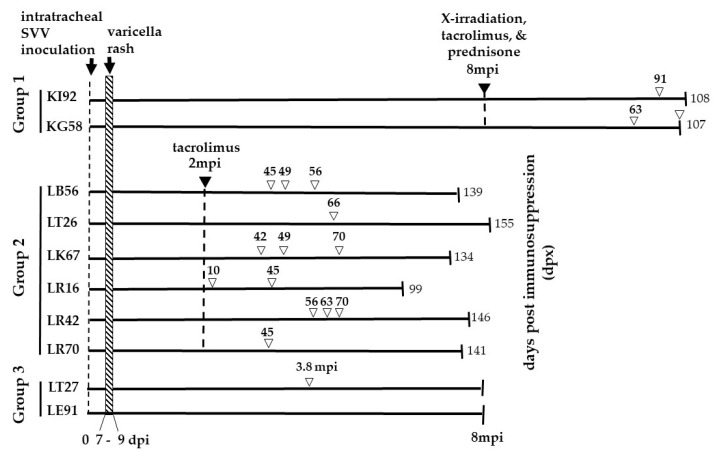
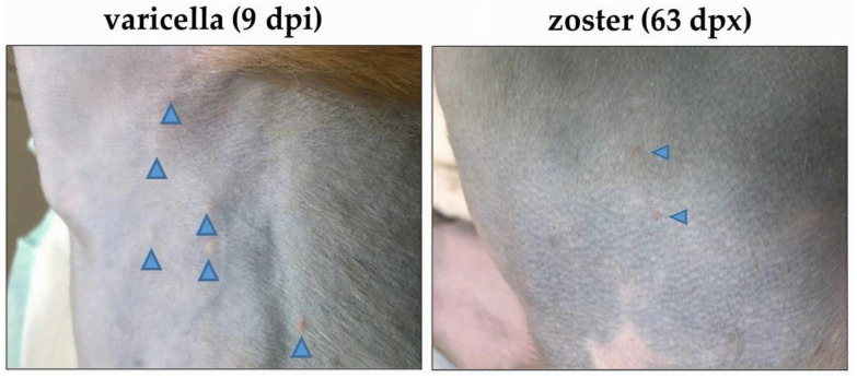
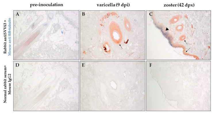
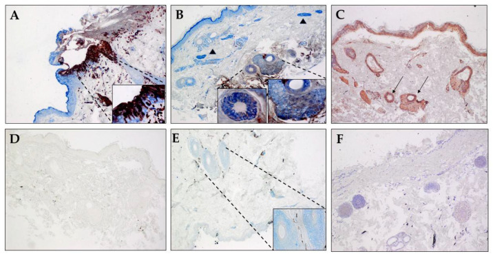
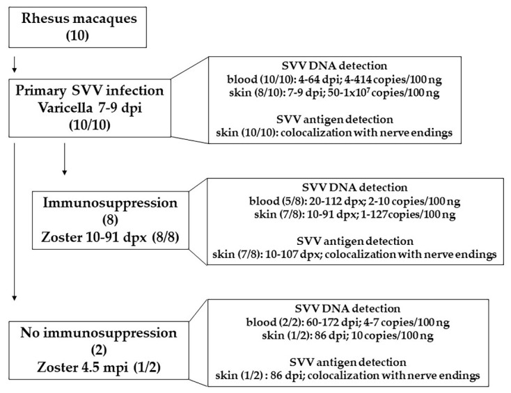
Primary simian varicella virus (SVV) infection and reactivation in nonhuman primates is a valuable animal model in the study of varicella zoster virus disease [varicella (chickenpox) and herpes zoster (shingles)]. To understand SVV pathogenesis in skin, we inoculated 10 rhesus macaques with SVV, resulting in varicella rash. After the establishment of latency, eight of the monkeys were immunosuppressed using tacrolimus with or without irradiation and prednisone and two monkeys were not immunosuppressed. Zoster rash developed in all immunosuppressed monkeys and in one non-immunosuppressed monkey. Five monkeys had recurrent zoster. During varicella and zoster, SVV DNA in skin scrapings ranged from 50 to 107 copies/100 ng of total DNA and 2-127 copies/100 ng of total DNA, respectively. Detection of SVV DNA in blood during varicella was more frequent and abundant compared to that of zoster. During varicella and zoster, SVV antigens colocalized with neurons expressing β-III tubulin in epidermis, hair follicles, and sweat glands, suggesting axonal transport of the virus. Together, we have demonstrated that both SVV DNA and antigens can be detected in skin lesions during varicella and zoster, providing the basis for further studies on SVV skin pathogenesis, including immune responses and mechanisms of peripheral spread.

Keywords: simian varicella virus; skin pathogenesis; varicella; zoster.


Conflict of interest statement

The authors declare no conflict of interest. The funders had no role in the design of the study; in the collection, analyses, or interpretation of data; in the writing of the manuscript, or in the decision to publish the results.

Figures

Figure 1
Figure 1
Experimental Design. The monkeys were divided into three groups. Two rhesus macaques (Group 1—KI92 and KG58) were intratracheally inoculated with 5 × 10 5 pfu of SVV and developed varicella rash 7–9 days post-inoculation (dpi). Eight months’ post-inoculation (mpi), both monkeys were exposed to X-irradiation (200 cGy) and daily oral treatment with tacrolimus (80 µg/kg/day) and prednisone (1 mg/kg/day) for the duration of the experiment. KI92 and KG58 developed zoster rash, 91- and 63-days post-immunosuppression treatment (dpx), respectively. KG58 had recurrent zoster at 107 dpx. KI92 and KG58 were euthanized 108 and 107 dpx, respectively. Six rhesus macaques (group 2—LB56, LT26, LK67, LR16, LR42, and LR70) were intratracheally inoculated with 2.4 × 10 5 pfu SVV, and all developed varicella rash between 7–9 dpi. Two months later, all six monkeys were treated with tacrolimus (80 µg/kg/day) daily for the duration of the experiment. All six immunosuppressed monkeys developed a zoster rash at indicated dpx (numbers above, unfilled arrowheads). Monkeys LB56, LK67, LR16, and LR42 had recurrent zoster, as indicated by multiple unfilled arrowheads. All six monkeys were euthanized at the indicated dpx. Two rhesus macaques (group 3—LT27 and LE91) were intratracheally inoculated with 2.4 × 10⁵ pfu of SVV, and both developed varicella rash by nine dpi. Neither were immunosuppressed. One of the monkeys (LT27) developed zoster rash 3.8 mpi. Group 3 monkeys were euthanized 8 mpi. DNA extracted from skin scrapings obtained at the time of zoster from monkeys in all three groups were analyzed for the presence of SVV DNA by qPCR. Euthanasia is indicated by vertical lines on the right end of the lines. The numbers at the end of the lines indicate the time (dpx (groups 1–2) or mpi (group 3)) of euthanasia.
Figure 2
Figure 2
Skin rash after varicella and zoster in monkey LR42 (group 2). Varicella skin rash was seen in the torso nine days after SVV inoculation (dpi; left image). Zoster skin rash was seen on the torso 63 days after immunosuppression (dpx) with tacrolimus treatment (right image). The location of the vesicles is indicated by blue arrowheads.
Figure 3
Figure 3
Immunohistochemical analysis of varicella and zoster skin rash from the same rhesus macaque. Sections of biopsied, zinc formalin-fixed, paraffin-embedded skin samples before inoculation with SVV (pre-inoculation), 9 dpi (days post-inoculation) and 42 dpx (days post-immunosuppression) from rhesus macaque LK67 (group 2) was analyzed using dual-color staining. Rabbit polyclonal antibodies against SVV IE63 protein (red) and mouse monoclonal antibody directed against human β-III tubulin (blue) were used as described in methods to identify the presence of virus antigen and its proximity to nerve endings. Normal rabbit serum and mouse anti IgG2a were used as negative controls (DF). SVV IE63 protein was not detected in the pre-inoculation biopsy (A), but was found in hair follicles during varicella and zoster and in the epidermis during zoster (B, arrows). β-III tubulin was found to colocalize with SVV IE63 protein (C, arrowhead) in the epidermis during zoster. (Magnification, ×100).
Figure 4
Figure 4
Colocalization of SVV antigen and βIII-tubulin in skin sweat glands during zoster. Sections of biopsied, zinc-formalion-fixed, paraffin-embedded skin samples from (A) an acutely infected immunosuppressed rhesus macaque (B321; SVV-positive control), (BD) from rhesus macaque KI92 (group 1) 91 dpx (days post-immunosuppression), and (E,F) an uninfected SVV-seronegative rhesus macaque (R110368) were analyzed by immunohistochemistry. Rabbit polyclonal antibodies against SVV IE63 protein (red) (A,B,E), SVV gH + L, and (C) mouse monoclonal antibody directed against human β-III tubulin (blue; A,B,E) were used as described in methods. Nerve bundles containing βIII-tubulin are identified by arrowheads in panel B. Normal rabbit serum by itself (F) or with mouse anti IgG2a antibody (D) were used as negative controls. Colocalization of SVV IE63 protein with β-III tubulin can be seen in epidermis during acute infection (A, inset) and in sweat glands and hair follicles during reactivation (B, inset). SVV gH + L antigens can also be seen in sweat glands in an adjacent section (C, thin arrows). β-III tubulin, but not SVV IE63, protein can be seen in sweat glands and hair follicle in skin from an uninfected monkey (E, inset). Normal rabbit serum along with mouse anti IgG2a antibody on skin from zoster in monkey KI92 (D) or using rabbit anti SVV gH + L on skin section from the uninfected monkey (F) did not show any staining. Section in panel F was counterstained with hematoxylin. (Magnification, ×100, inset ×600).
Figure 5
Figure 5
Summary of findings in this report. Ten rhesus macaques were inoculated with SVV. All of them developed varicella 7–9 dpi. SVV DNA (4–414 copies/100 ng) was detected in blood, 4–64 days post infection (dpi) in all 10 monkeys. SVV DNA (50–1 × 10⁷ copies/100 ng) was detected in skin scrapings in 8 of 10 monkeys, 7–9 dpi. Analysis of biopsied skin samples, during varicella, revealed colocalization of SVV antigens with nerve endings in all 10 monkeys. Establishment of latency was confirmed by the absence of SVV DNA in blood. Eight of latently infected monkeys were immunosuppressed. Zoster rash developed in all monkeys 10–91 days post immunosuppression (dpx). SVV DNA (2–10 copies/100 ng) was detected in blood, 20–112 dpx, in 5 of 8 monkeys. During zoster (10–91 dpx), 1–127 copies of SVV DNA (per 100 ng) were detected in skin scrapings. In the affected skin, SVV antigens were found to be colocalized with nerve endings. In two monkeys were not immunosuppressed, SVV DNA (4–7 copies/100 ng) was detected 60–172 dpi. One of the two non-immunosuppressed monkey developed zoster, 4.5 mpi (months post infection). SVV DNA (10 copies/100 ng) and antigens was detected in skin scrapings during zoster (86 dpi). SVV antigen was found to be colocalized with nerve endings. Abundance of SVV DNA and antigens in skin was substantially higher during varicella compared to zoster. SVV infection and reactivation in rhesus macaques serves as an extremely useful model to study varicella and zoster in humans.

References

    1. Ku C.-C., Zerboni L., Ito H., Graham B.S., Wallace M., Arvin A.M. Varicella-Zoster Virus Transfer to Skin by T Cells and Modulation of Viral Replication by Epidermal Cell Interferon-α. J. Exp. Med. 2004;200:917–925. doi: 10.1084/jem.20040634. - DOI - PMC - PubMed
    1. Kimura H., Kido S., Ozaki T., Tanaka N., Ito Y., Williams R.K., Morishima T. Comparison of quantitations of viral load in varicella and zoster. J. Clin. Microbiol. 2000;38:2447–2449. doi: 10.1128/JCM.38.6.2447-2449.2000. - DOI - PMC - PubMed
    1. Ishizaki Y., Tezuka J., Ohga S., Nomura A., Suga N., Kuromaru R., Kusuhara K., Mizuno Y., Kasuga N., Hara T. Quantification of circulating varicella zoster virus-DNA for the early diagnosis of visceral varicella. J. Infect. 2003;47:133–138. doi: 10.1016/S0163-4453(03)00004-5. - DOI - PubMed
    1. Kronenberg A., Bossart W., Wuthrich R.P., Cao C., Lautenschlager S., Wiegand N.D., Mullhaupt B., Noll G., Mueller N.J., Speck R.F. Retrospective analysis of varicella zoster virus (VZV) copy DNA numbers in plasma of immunocompetent patients with herpes zoster, of immunocompromised patients with disseminated VZV disease, and of asymptomatic solid organ transplant recipients. Transpl. Infect. Dis. 2005;7:116–121. doi: 10.1111/j.1399-3062.2005.00106.x. - DOI - PubMed
    1. Satyaprakash A.K., Tremaine A.M., Stelter A.A., Creed R., Ravanfar P., Mendoza N., Mehta S.K., Rady P.L., Pierson D.L., Tyring S.K. Viremia in Acute Herpes Zoster. J. Infect. Dis. 2009;200:26–32. doi: 10.1086/599381. - DOI - PubMed

Publication types