. 2022 Jun 5;14(6):1225.
doi: 10.3390/v14061225.

Characterization of the Cross-Species Transmission Potential for Porcine Deltacoronaviruses Expressing Sparrow Coronavirus Spike Protein in Commercial Poultry

Affiliations

Characterization of the Cross-Species Transmission Potential for Porcine Deltacoronaviruses Expressing Sparrow Coronavirus Spike Protein in Commercial Poultry

Moyasar A Alhamo et al. Viruses. .

Abstract

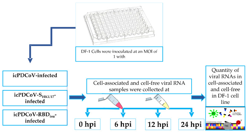
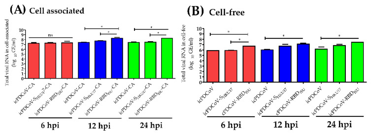
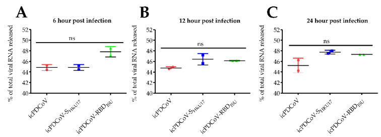
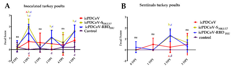
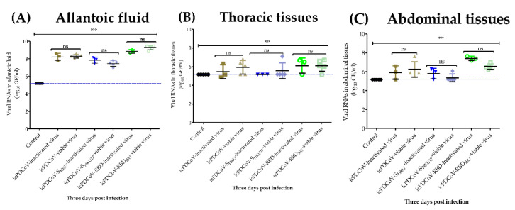
Avian species often serve as transmission vectors and sources of recombination for viral infections due to their ability to travel vast distances and their gregarious behaviors. Recently a novel deltacoronavirus (DCoV) was identified in sparrows. Sparrow deltacoronavirus (SpDCoV), coupled with close contact between sparrows and swine carrying porcine deltacoronavirus (PDCoV) may facilitate recombination of DCoVs resulting in novel CoV variants. We hypothesized that the spike (S) protein or receptor-binding domain (RBD) from sparrow coronaviruses (SpCoVs) may enhance infection in poultry. We used recombinant chimeric viruses, which express S protein or the RBD of SpCoV (icPDCoV-SHKU17, and icPDCoV-RBDISU) on the genomic backbone of an infectious clone of PDCoV (icPDCoV). Chimeric viruses were utilized to infect chicken derived DF-1 cells, turkey poults, and embryonated chicken eggs (ECEs) to examine permissiveness, viral replication kinetics, pathogenesis and pathology. We demonstrated that DF-1 cells in addition to the positive control LLC-PK1 cells are susceptible to SpCoV spike- and RBD- recombinant chimeric virus infections. However, the replication of chimeric viruses in DF-1 cells, but not LLC-PK1 cells, was inefficient. Inoculated 8-day-old turkey poults appeared resistant to icPDCoV-, icPDCoV-SHKU17- and icPDCoV-RBDISU virus infections. In 5-day-old ECEs, significant mortality was observed in PDCoV inoculated eggs with less in the spike chimeras, while in 11-day-old ECEs there was no evidence of viral replication, suggesting that PDCoV is better adapted to cross species infection and differentiated ECE cells are not susceptible to PDCoV infection. Collectively, we demonstrate that the SpCoV chimeric viruses are not more infectious in turkeys, nor ECEs than wild type PDCoV. Therefore, understanding the cell and host factors that contribute to resistance to PDCoV and avian-swine chimeric virus infections may aid in the design of novel antiviral therapies against DCoVs.

Keywords: S protein; chicken embryos; coronaviruses; cross-species infection; porcine delta coronavirus; sparrow delta coronavirus; turkey poults.


Conflict of interest statement

The authors declare no conflict of interest.

Figures

Figure 1
Figure 1
Schematic diagram of the DF-1 cells showing treatment groups and time points at which cell-associated and cell-free were collected for viral RNA titration.
Figure 2
Figure 2
Schematic diagram of the experimental design showing treatment groups and time points at which the sentinels were commingled, and the birds were euthanized. Turkey poults at 8 days of age were infected and euthanized at 11 and 13 days of age. Sentinel birds at 10 days of age accordingly commingled with each infected group and euthanized at 12 days of age. Turkey poults inoculated with 4.4 × 10⁵ FFU/poult of icPDCoV-(passage 8), icPDCoV-SHKU17-(passage 3), and icPDCoV-RBDISU (passage 3), viruses, or MEM in a volume of 200 µL. DPC, day post commingling.
Figure 3
Figure 3
Indirect immunofluorescence imaging of PDCoV N antigen in DF-1 and LLC-PK1 cell lines. Cell lines were inoculated with mock inoculated (A,B), or inoculated with icPDCoV (C,D), icPDCoV-SHKU17 (E,F), and icPDCoV-RBDISU (G,H) viruses at an MOI of 0.01. DF-1 and LLC-PK1 cells were fixed at 18 hpi or 12 hpi and stained using a mouse monoclonal, SD55-197 against PDCoV N protein, respectively. Representative images are shown (labeling for PDCoV N antigen in the infected cells (in green) and nuclei (in blue)). Scale bara of the zoom: 1.5 are 22.5.
Figure 4
Figure 4
Replication kinetics of icPDCoV, icPDCoV-SHKU17, and icPDCoV-RBDISU viruses. (A) LLC-PK1 and DF-1 cell lines were inoculated at an MOI of 0.01 (4A) and DF-1 cells at an MOI of 10 (4B), respectively. LLC-PK1 kinetics at 0.01 MOI was reported previously by Niu et al. [31]. The data for icPDCoV were included in 4A as a TCID50 kinetics reference. After 1 h of incubation at 37 °C, cells were washed and overlaid with MEM supplemented with 10 µg/mL trypsin (LLC-PK-1) or no trypsin (DF-1) due to cell sensitivity. The kinetics of viral replication were evaluated in supernatants collected at denoted timepoints post infection, and the viral titers were determined by TCID50 (A,B). Different lower-case letters (a, b, c) indicate significant differences between each group (n = 3 biologically independent samples) at each time point. All data points are mean ± SD. Two-way ANOVA with Bonferroni post hoc test, p ≤ 0.05. ns, not statistically significant. (CE) Light microscopy of DF-1 cells infected with specified virus at 72 h post infection with an MOI of 0.01.
Figure 5
Figure 5
Comparison of viral RNA loads in inoculated DF-1 cells. The DF-1 cells were inoculated with icPDCoV, icPDCoV-SHKU17, and icPDCoV-RBDISU viruses at an MOI of 1. Data shown are levels of virus quantified in DF-1 from virus associated with DF-1 compared with the same inoculated DF-1 cells in cell-free fraction at (A) 0 hpi, (B) 6 hpi and (C) 12 hpi, and (D) 24 hpi. The cutoff value was 5.17 log10 GE/mL. hpi, hour post infection; GE, genomic equivalent; CA, cell-associated; CF, cell-free. One-way ANOVA with Tukey’s multiple comparisons post hoc test, ** p ≤ 0.05, ns; not statistically significant.
Figure 6
Figure 6
Comparison of viral RNA loads in virus associated with DF-1 and cell-free supernatant. The DF-1 cells were inoculated with icPDCoV, icPDCoV-SHKU17, and icPDCoV-RBDISU viruses at an MOI of 1. Data shown are levels of virus quantified in DF-1 from virus associated with DF-1 compared with the same inoculated DF-1 cells in cell-free fraction at 6 hpi, 12 hpi, and 24 hpi. hpi, hour post infection; GE, genomic equivalent; CA, cell-associated; CF, cell-free. One-way ANOVA with Tukey’s multiple comparisons post hoc test, * p ≤ 0.05, ns; not statistically significant.
Figure 7
Figure 7
Released ratios of viral RNAs in inoculated DF-1 cells. The DF-1 cells were inoculated with icPDCoV, icPDCoV-SHKU17, and icPDCoV-RBDISU viruses at an MOI of 1. Data shown are levels of virus quantified in DF-1 from virus associated with DF-1 compared with the same inoculated DF-1 cells in cell-free fraction at (A) 6 hpi, (B) 12 hpi, and (C) 24 hpi. All data points (cts) were first converted to GE/mL, and then, the percentage of releases was calculated, according to the equation previously described. GE, genomic equivalent. One-way ANOVA with Tukey’s multiple comparisons post hoc test, p ≤ 0.05, ns; not statistically significant.
Figure 8
Figure 8
Fecal scores of turkey poults at 0- to 5-DPI. Fecal consistency was scored as follows: 0, normal; 1, pasty; 2, semiliquid; 3, watery, and the fecal score >2 was considered as diarrhea. Red circles, green squares, blue triangles, and black triangles represent (A) icPDCoV-inoculated, icPDCoV-SHKU17-inoculated, icPDCoV-RBDISU-inoculated, and control groups (n = 10 biologically independent samples). (B) sentinel poults that commingled at 1 DPI (n = 4 biologically independent samples), respectively. Different lower-case letters (a; icPDCoV, b; icPDCoV-SHKU17, c; icPDCoV-RBDISU, d; mock) indicate significant differences between the given group at each time point. Significant diarrhea for each test virus was only observed at day 1 post infection but for groups icPDCoV-SHKU17 and icPDCoV-RBDISU diarrhea was also observed on days 3 and 5 in inoculated birds and day 2 in sentinel birds. Two-way ANOVA with Bonferroni post hoc test, p ≤ 0.05. ns, not statistically significant.
Figure 9
Figure 9
Immunohistochemistry was used to detect PDCoV NP in the lung, duodenum, jejunum, or ileum tissues from turkey poults. Birds were inoculated with- icPDCoV (A,D,G,J), icPDCoV-SHKU17 (B,E,H,K), or icPDCoV-RBDISU (C,F,I,L), showing no cellular signal for PDCoV N antigen. PDCoV-infected pig intestine used as positive (M) and negative (N) controls (magnification, 300×).
Figure 10
Figure 10
Quantity of viral RNAs in (A) allantoic fluids, (B) thoracic- and (C) abdominal tissues. The 11-day-old embryonated chicken eggs were inoculated with mock-inoculated, or heat-inactivated (at 60 °C for 20 min) or viable icPDCoV, icPDCoV-SHKU17, and icPDCoV-RBDISU viruses. Dashed line indicates detection limit of 5.17 log10 GE/mL of viruses in samples. GE, genomic equivalent. One-way ANOVA with Tukey’s multiple comparisons post hoc test, *** p ≤ 0.05, ns; not statistically significant.

Similar articles

Cited by

References

    1. Wang Q., Vlasova A.N., Kenney S.P., Saif L.J. Emerging and re-emerging coronaviruses in pigs. Curr. Opin. Virol. 2019;34:39–49. doi: 10.1016/j.coviro.2018.12.001. - DOI - PMC - PubMed
    1. Cui J., Li F., Shi Z.-L. Origin and evolution of pathogenic coronaviruses. Nat. Rev. Microbiol. 2019;17:181–192. doi: 10.1038/s41579-018-0118-9. - DOI - PMC - PubMed
    1. Masters P.S., Perlman S. In: Fields Virology. Knipe D.M., Howley P.M., editors. Vol. 2. Lippincott Williams & Wilkins; Philadelphia, PA, USA: 2013. pp. 825–858.
    1. Kenney S.P., Wang Q., Vlasova A., Jung K., Saif L. Naturally Occurring Animal Coronaviruses as Models for Studying Highly Pathogenic Human Coronaviral Disease. Veter. Pathol. 2021;58:438–452. doi: 10.1177/0300985820980842. - DOI - PubMed
    1. Ksiazek T.G., Erdman D., Goldsmith C.S., Zaki S.R., Peret T., Emery S., Tong S., Urbani C., Comer J.A., Lim W., et al. A Novel Coronavirus Associated with Severe Acute Respiratory Syndrome. N. Engl. J. Med. 2003;348:1953–1966. doi: 10.1056/NEJMoa030781. - DOI - PubMed

Publication types

Substances

Supplementary concepts

LinkOut - more resources