

Antiviral Activities of Green Tea Components against Grouper Iridovirus Infection In Vitro and In Vivo
- PMID: 35746698
- PMCID: PMC9227864
- DOI: 10.3390/v14061227
Antiviral Activities of Green Tea Components against Grouper Iridovirus Infection In Vitro and In Vivo
Abstract
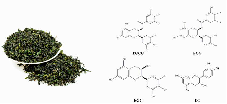
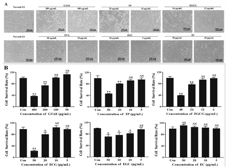
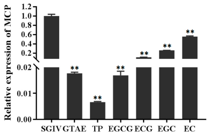
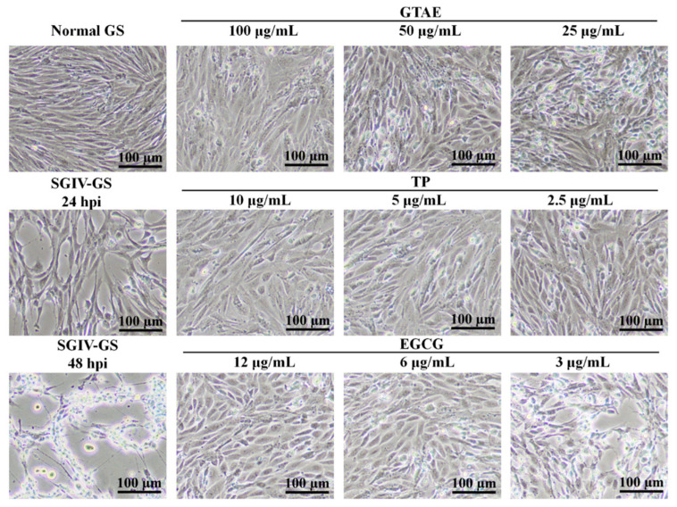
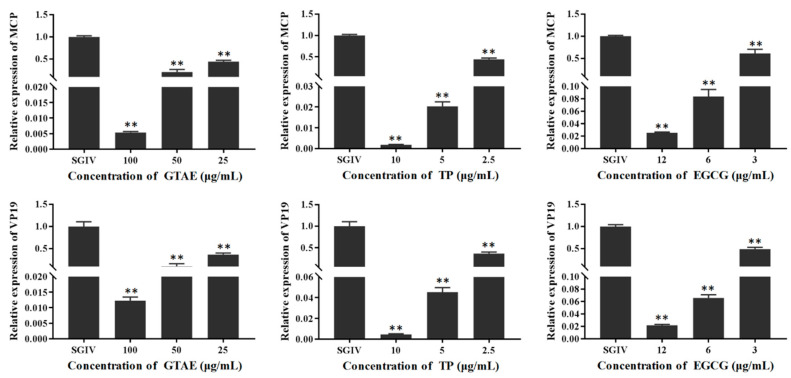
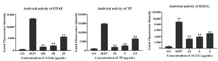
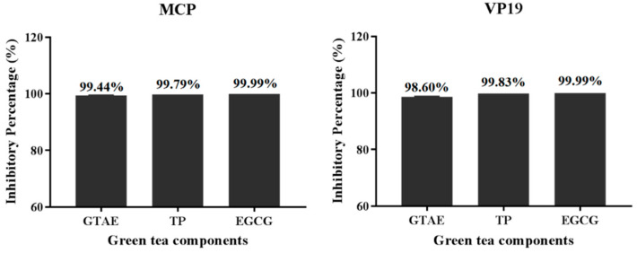
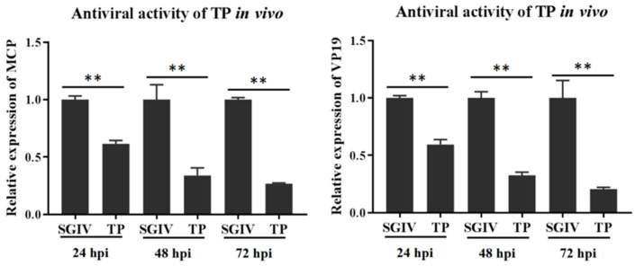
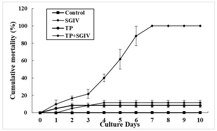
(1) Background: Singapore grouper iridovirus (SGIV) can cause extensive fish deaths. Therefore, developing treatments to combat virulent SGIV is of great economic importance to address this challenge to the grouper aquaculture industry. Green tea is an important medicinal and edible plant throughout the world. In this study, we evaluated the use of green tea components against SGIV infection. (2) Methods: The safe working concentrations of green tea components were identified by cell viability detection and light microscopy. Additionally, the antiviral activity of each green tea component against SGIV infection was determined with light microscopy, an aptamer (Q5c)-based fluorescent molecular probe, and reverse transcription quantitative PCR. (3) Results: The safe working concentrations of green tea components were green tea aqueous extract (GTAE) ≤ 100 μg/mL, green tea polyphenols (TP) ≤ 10 μg/mL, epigallocatechin-3-gallate (EGCG) ≤ 12 μg/mL, (-)-epigallocatechin (EGC) ≤ 10 μg/mL, (-)-epicatechin gallate (EGC) ≤ 5 μg/mL, and (-)-epicatechin (EC) ≤ 50 μg/mL. The relative antiviral activities of the green tea components determined in terms of MCP gene expression were TP > EGCG > GTAE > ECG > EGC > EC, with inhibition rates of 99.34%, 98.31%, 98.23%, 88.62%, 73.80%, and 44.31%, respectively. The antiviral effect of aptamer-Q5c was consistent with the results of qPCR. Also, TP had an excellent antiviral effect in vitro, wherein the mortality of fish in only the SGIV-injection group and TP + SGIV-injection group were 100% and 11.67%, respectively. (4) Conclusions: In conclusion, our results suggest that green tea components have effective antiviral properties against SGIV and may be candidate agents for the effective treatment and control of SGIV infections in grouper aquaculture.
Keywords: EGCG; antiviral activity; green tea component; grouper iridovirus; tea polyphenol.
Conflict of interest statement
The authors declare no conflict of interest.
Figures
Similar articles
-
Effect of EGCG Extracted from Green Tea against Largemouth Bass Virus Infection.Viruses. 2023 Jan 3;15(1):151. doi: 10.3390/v15010151. Viruses. 2023. PMID: 36680191 Free PMC article.
-
Antiviral activity of epicatechin against Singapore grouper iridovirus in vitro and in vivo.Fish Shellfish Immunol. 2025 Jul;162:110331. doi: 10.1016/j.fsi.2025.110331. Epub 2025 Apr 11. Fish Shellfish Immunol. 2025. PMID: 40222693
-
Research on the indirect antiviral function of medicinal plant ingredient quercetin against grouper iridovirus infection.Fish Shellfish Immunol. 2022 May;124:372-379. doi: 10.1016/j.fsi.2022.04.013. Epub 2022 Apr 14. Fish Shellfish Immunol. 2022. PMID: 35430348
-
Antiviral Effect and Mechanism of Edaravone against Grouper Iridovirus Infection.Viruses. 2023 Nov 10;15(11):2237. doi: 10.3390/v15112237. Viruses. 2023. PMID: 38005914 Free PMC article.
-
The medicinal value of tea drinking in the management of COVID-19.Heliyon. 2023 Jan;9(1):e12968. doi: 10.1016/j.heliyon.2023.e12968. Epub 2023 Jan 12. Heliyon. 2023. PMID: 36647394 Free PMC article. Review.
Cited by
-
Effect of EGCG Extracted from Green Tea against Largemouth Bass Virus Infection.Viruses. 2023 Jan 3;15(1):151. doi: 10.3390/v15010151. Viruses. 2023. PMID: 36680191 Free PMC article.
-
Exploring the Therapeutic Potential of Green Tea (Camellia sinensis L.) in Anti-Aging: A Comprehensive Review of Mechanisms and Findings.Mini Rev Med Chem. 2025;25(5):403-424. doi: 10.2174/0113895575331878240924035332. Mini Rev Med Chem. 2025. PMID: 39377377 Review.
-
Antimicrobial and antioxidant activity of encapsulated tea polyphenols in chitosan/alginate-coated zein nanoparticles: a possible supplement against fish pathogens in aquaculture.Environ Sci Pollut Res Int. 2024 Feb;31(9):13673-13687. doi: 10.1007/s11356-024-32058-x. Epub 2024 Jan 23. Environ Sci Pollut Res Int. 2024. PMID: 38261222 Free PMC article.
-
Epigallocatechin-3-gallate Attenuates the Bromo-3-chloro-5,5-dimethylhydantoin-induced Immunotoxicity in Crayfish.Mar Biotechnol (NY). 2025 Mar 27;27(2):69. doi: 10.1007/s10126-025-10449-6. Mar Biotechnol (NY). 2025. PMID: 40146333
References
-
- Li P., Wei S., Zhou L., Yang M., Yu Y., Wei J., Jiang G., Qin Q. Selection and characterization of novel DNA aptamers specifically recognized by Singapore grouper iridovirus-infected fish cells. J. Gen. Virol. 2015;96:3348–3359. - PubMed
-
- Yu Q., Liu M.Z., Wei S.N., Xiao H.H., Wu S.T., Qin X.L., Shi D.Q., Li S.Q., Wang T.X., Li P.F. Isolation of Nervous Necrosis Virus from Hybrid Grouper (Epinephelus fuscoguttatus♀ × Epinephelus lanceolatus♂) Cultured in Guangxi, China. Fish Pathol. 2019;54:16–19. doi: 10.3147/jsfp.54.16. - DOI
Publication types
MeSH terms
Substances
LinkOut - more resources
Full Text Sources
Miscellaneous
