. 2022 Jun 7;14(6):1233.
doi: 10.3390/v14061233.

Interplay of HCPro and CP in the Regulation of Potato Virus A RNA Expression and Encapsidation

Affiliations

Interplay of HCPro and CP in the Regulation of Potato Virus A RNA Expression and Encapsidation

Shreya Saha et al. Viruses. .

Abstract

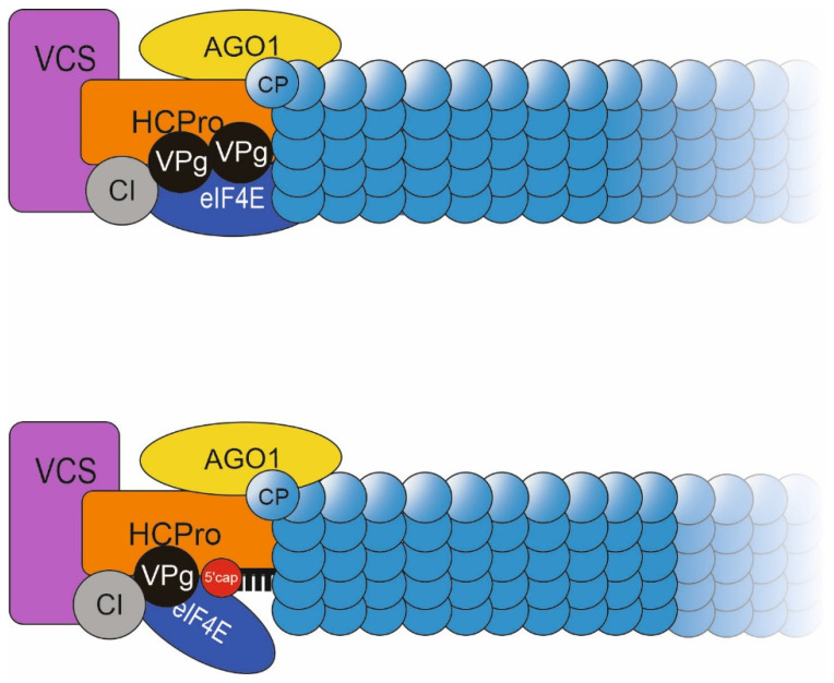
Potyviral coat protein (CP) and helper component-proteinase (HCPro) play key roles in both the regulation of viral gene expression and the formation of viral particles. We investigated the interplay between CP and HCPro during these viral processes. While the endogenous HCPro and a heterologous viral suppressor of gene silencing both complemented HCPro-less potato virus A (PVA) expression, CP stabilization connected to particle formation could be complemented only by the cognate PVA HCPro. We found that HCPro relieves CP-mediated inhibition of PVA RNA expression likely by enabling HCPro-mediated sequestration of CPs to particles. We addressed the question about the role of replication in formation of PVA particles and gained evidence for encapsidation of non-replicating PVA RNA. The extreme instability of these particles substantiates the need for replication in the formation of stable particles. During replication, viral protein genome linked (VPg) becomes covalently attached to PVA RNA and can attract HCPro, cylindrical inclusion protein and host proteins. Based on the results of the current study and our previous findings we propose a model in which a large ribonucleoprotein complex formed around VPg at one end of PVA particles is essential for their integrity.

Keywords: coat protein (CP); encapsidation; helper-component proteinase (HCPro); particle stability; potato virus A; potyvirus; viral gene expression.


Conflict of interest statement

The authors declare no conflict of interest.

Figures

Figure 1
Figure 1
Schematic diagrams of viral constructs used in this study.
Figure 2
Figure 2
PVA HCPro alone does not increase CP stability in plant tissue lysate. (A) PVA CP was co-expressed with RFP-tagged versions of either PVA HCPro, HCProWD, HCPro4Ebd or HCProSD, and the samples for western blot analysis were collected at 4 dpi. Neither HCPro nor its mutants were able to stabilize PVA CP in the in vitro degradation assay. Below is the loading control, Rubisco large subunit (RbcL) band stained with Ponceau S. (B) Both PVA HCProRFP and CPYFP localized all over the cytoplasm when they were co-expressed. Given is also the overlay of the CPYFP and HCProRFP fluorescence.
Figure 3
Figure 3
Transiently expressed HCPro complements gene expression and virion formation of HCPro-less PVA. (A) The RLUC activity was measured from PVAΔHCPro co-expressed with the GUS control, HCPro, HCProWD or cucumoviral suppressor of RNA silencing, 2b. (B) Total RNA of PVAΔHCPro in the same experiment as in (A). (C) Quantitation of PVAΔHCPro RNA by RT-qPCR of immuno-captured templates in the same experiment as in (A). All the samples in (AC) were collected at 5 dpi. RLUC, total RNA and particle-associated RNA levels from PVAWT infected plants is shown for comparison. (D) Samples from this experiment were subjected to in vitro degradation assay. A western blot analysis with anti-CP antibodies of the samples as they were at 0 min time point and after 60 min incubation of the plant sap at room temperature. RbcL band in Ponceau S-stained membrane is given as a loading control. p < 0.05 = *; p < 0.01 = **.
Figure 4
Figure 4
PVA CP inhibits viral RNA translation from the entire length of PVA RNA. (A) Viral translation from PVAWT carrying the rluc cistron in front of the HCPro cistron (PVAWT:RLUCH) was determined as RLUC activity at 3 dpi. (B) The western blot analyses with anti-CP antibodies show CP production from the PVA genome in the GUS control lane and its transient overexpression in the CP lane. Notably, PVA RNA translation is blocked in the latter case. p < 0.01 = **.
Figure 5
Figure 5
HCPro relieves the CP-mediated translational block of PVA. (A) PVAWT was co-expressed with GUS, CP or HCPro alone or with CP and HCPro together. RLUC and CP levels were analyzed at 3 dpi. (B) Same as (A) but with the PVAΔGDD. (C) Same as (A) but with PVACPmut. (D) PVAWT RNA amount from virions was measured by IC-RT-qPCR at 9 dpi in the same setup as in (A) p < 0.05 = *; p < 0.01 = **; p < 0.001 = ***. (E,F) Western blots showing CP expression in (A,D), respectively.
Figure 6
Figure 6
IC-RT-qPCR detects little replication-defective RNA. (A) PVAWT and PVAWD alone and PVAΔGDD together with either GUS, CP, or CP plus HCPro were transiently expressed in N. bet-hamiana leaves. Gene expression from PVAWT, PVAWD and PVAΔGDD was quantitated at 5 dpi. The graph is presented in logarithmic scale to accommodate the vast differences in the gene expression amongthe mutant virus infected samples. (B) Quantitation of the total genomic vRNA amounts of the same samples as in (A). (C) Amount of genomic vRNA detected in the same samples as in (B) with IC-RT-qPCR indicating the amount of encapsidated PVA RNA. p < 0.01 = **, p < 0.05 = *.
Figure 7
Figure 7
Degradation-prone particles are formed both by PVAΔGDD and PVAWD. (A) In vivo cross-linking of PVA particles. RNA copy numbers for the PVAWT, PVAΔGDD + HCPro + CP, PVAΔGDD, PVAWD detected from samples derived from non-fixed tissue and fixed tissue after reversion of the cross-link (marked by F). Student’s t-test between the non-fixed and fixed samples of each construct type was carried out (p < 0.05 = *). (B) PVAWT particles and virus-like particles from PVAΔGDD + HCPro + CP, PVAΔGDD and PVAWD visualized with negative staining and transmission electron microscopy. Arrows indicate representative particles. (C) Particle size measurements. Fixed PVAWT, PVAΔGDD, PVAWD particles and particle-like structures detected in TEM images were measured and the values are presented in boxplots.
Figure 8
Figure 8
Virus particles were not detected in thin sections of PVAΔGDD-infected tissues. (A) Virus particle stacks marked with black arrow heads were abundant in a PVAWT infected leaves but were not observed in PVAΔGDD samples at 8 dpi. PVAWT and PVAΔGDD were infiltrated at OD600 0.1 and 0.2. PVAWT was supplemented with GUS at OD600 0.6 and PVAΔGDD with HCPro and CP both at OD600 0.3. Samples were taken from the infiltrated leaves at 8 dpi and fixed in 2.5% glutaraldehyde prior to visualization by thin-section transmission electron microscopy. (B,C) Transient expression of CP and HCPro in PVAΔGDD + CP + HCPro leaves was confirmed by anti-CP and anti-HCPro western blots, respectively. Gel loading was checked by staining with Ponceau solution.
Figure 9
Figure 9
A hypothetical model for the formation of stable PVA particles. (Top): PVAWT particle with a high-molecular weight ribonucleoprotein complex assembled around VPg covalently bound at the 5′ end of vRNA. The model incorporates known interactions between HCPro and VPg, CI, eiIF(iso)4E, VCS and AGO1 and CP. (Bottom): A PVAΔGDD-derived particle where the m7G cap replaces the covalently-bound VPg at the 5′ end thus leaving the vRNA exposed to host RNases.

References

    1. Valli A.A., Gallo A., Rodamilans B., López-Moya J.J., García J.A. The HCPro from the potyviridae family: An enviable multitasking helper component that every virus would like to have. Mol. Plant Pathol. 2018;19:744–763. doi: 10.1111/mpp.12553. - DOI - PMC - PubMed
    1. Anandalakshmi R., Pruss G.J., Ge X., Marathe R., Mallory A.C., Smith T.H., Vance V.B. A viral suppressor of gene silencing in plants. Proc. Natl. Acad. Sci. USA. 1998;95:13079–13084. doi: 10.1073/pnas.95.22.13079. - DOI - PMC - PubMed
    1. Kasschau K.D., Carrington J.C. A counterdefensive strategy of plant viruses: Suppression of posttranscriptional gene silencing. Cell. 1998;95:461–470. doi: 10.1016/S0092-8674(00)81614-1. - DOI - PubMed
    1. González-Jara P., Atencio F.A., Martínez-García B., Barajas D., Tenllado F., Díaz-Ruíz J.R. A single amino acid mutation in the plum pox virus helper component-proteinase gene abolishes both synergistic and RNA silencing suppression activities. Phytopathology. 2005;95:894–901. doi: 10.1094/PHYTO-95-0894. - DOI - PubMed
    1. Shi X.M., Miller H., Verchot J., Carrington J.C., Vance V.B. Mutations in the region encoding the central domain of helper component-proteinase (HC-Pro) eliminate potato virus X/potyviral synergism. Virology. 1997;231:35–42. doi: 10.1006/viro.1997.8488. - DOI - PubMed

Publication types

Supplementary concepts

LinkOut - more resources