. 2022 Jun 9;14(6):1249.
doi: 10.3390/v14061249.

Serum Neutralizing and Enhancing Effects on African Swine Fever Virus Infectivity in Adherent Pig PBMC

Affiliations

Serum Neutralizing and Enhancing Effects on African Swine Fever Virus Infectivity in Adherent Pig PBMC

Jessica A Canter et al. Viruses. .

Abstract

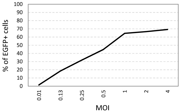
African swine fever virus (ASFV) causes hemorrhagic fever with mortality rates of up to 100% in domestic pigs. Currently, there are no commercial vaccines for the disease. Only some live-attenuated viruses have been able to protect pigs from ASFV infection. The immune mechanisms involved in the protection are unclear. Immune sera can neutralize ASFV but incompletely. The mechanisms involved are not fully understood. Currently, there is no standardized protocol for ASFV neutralization assays. In this study, a flow cytometry-based ASFV neutralization assay was developed and tested in pig adherent PBMC using a virulent ASFV containing a fluorescent protein gene as a substrate for neutralization. As with previous studies, the percentage of infected macrophages was approximately five time higher than that of infected monocytes, and nearly all infected cells displayed no staining with anti-CD16 antibodies. Sera from naïve pigs and pigs immunized with a live-attenuated ASFV and fully protected against parental virus were used in the assay. The sera displayed incomplete neutralization with MOI-dependent neutralizing efficacies. Extracellular, but not intracellular, virions suspended in naïve serum were more infectious than those in the culture medium, as reported for some enveloped viruses, suggesting a novel mechanism of ASFV infection in macrophages. Both the intracellular and extracellular virions could not be completely neutralized.

Keywords: African swine fever virus (ASFV); extracellular virions; flow cytometry; hyperimmune serum; monocyte-derived macrophage; serum-enhanced virus infection; virus neutralization.


Conflict of interest statement

The authors declare no conflict of interests.

Figures

Figure 1
Figure 1
The percentages of ASFV-infected (EGFP+) cells at different MOI (HAD50) in cultured pig ex vivo adherent PBMC, calculated based on flow cytometry analysis gated on the population of monocytes and macrophages.
Figure 2
Figure 2
The percentages of EGFP+ monocytes and macrophages infected with 200 μL of supernatants harvested at different hours post-infection from pig ex vivo adherent PBMC cultured in a 12-well plate and infected with MOI of 1 HAD50 (the percentages were calculated based on flow cytometry analysis gated on the population containing monocytes and macrophages).
Figure 3
Figure 3
The cell distribution on the SSC vs. FITC (EGFP signal) plot of adherent PBMC gated on monocytes and macrophages (red) and EGFP+ cells (green) after infection with ASFV at MOI of 0.5 HAD50 treated with (A) naïve serum (−) and (B) hyperimmune serum (+).
Figure 4
Figure 4
The neutralizing effects (percentage of EGFP+ cell reduction) of hyperimmunized sera compared to naïve sera or cell culture medium at MOI of 0.5 HAD50 (infection inoculum: 50 μL of ASFV in 250 μL of serum or medium) using adherent PBMC isolated from a donor pig.
Figure 5
Figure 5
The neutralization expressed as the percentages of protection of 2-fold serially diluted positive sera compared to 2-fold diluted negative sera [protection % = (PI − PI+)/PI, where PI is the percentage of infected cells treated with negative serum and PI+ is the percentage of infected cells treated with serum (−)] in gated monocytes and macrophages infected with different MOI.

Similar articles

Cited by

References

    1. Salguero F.J. Comparative pathology and pathogenesis of African swine fever infection in swine. Front. Vet. Sci. 2020;7:282. doi: 10.3389/fvets.2020.00282. - DOI - PMC - PubMed
    1. Gaudreault N.N., Richt J.A. Subunit vaccine approaches for African swine fever virus. Vaccines. 2019;7:56. doi: 10.3390/vaccines7020056. - DOI - PMC - PubMed
    1. Rock D.L. Thoughts on African swine fever vaccines. Viruses. 2021;13:943. doi: 10.3390/v13050943. - DOI - PMC - PubMed
    1. Onisk D.V., Borca M.V., Kutish G., Kramer E., Irusta P., Rock D.L. Passively transferred African swine fever virus antibodies protect swine against lethal infection. Virology. 1994;198:350–354. doi: 10.1006/viro.1994.1040. - DOI - PubMed
    1. Takamatsu H.H., Denyer M.S., Lacasta A., Stirling C.M., Argilaguet J.M., Netherton C.L., Oura C.A., Martins C., Rodríguez F. Cellular immunity in ASFV responses. Virus Res. 2013;173:110–121. doi: 10.1016/j.virusres.2012.11.009. - DOI - PubMed

Publication types