. 2022 Jun 14;14(6):1301.
doi: 10.3390/v14061301.

Photodynamic Inactivation of SARS-CoV-2 Infectivity and Antiviral Treatment Effects In Vitro

Affiliations

Photodynamic Inactivation of SARS-CoV-2 Infectivity and Antiviral Treatment Effects In Vitro

Svitlana Ziganshyna et al. Viruses. .

Abstract

Despite available vaccines, antibodies and antiviral agents, the severe acute respiratory syndrome coronavirus-2 (SARS-CoV-2) pandemic still continues to cause severe disease and death. Current treatment options are limited, and emerging new mutations are a challenge. Thus, novel treatments and measures for prevention of viral infections are urgently required. Photodynamic inactivation (PDI) is a potential treatment for infections by a broad variety of critical pathogens, including viruses. We explored the infectiousness of clinical SARS-CoV-2 isolates in Vero cell cultures after PDI-treatment, using the photosensitizer Tetrahydroporphyrin-tetratosylate (THPTS) and near-infrared light. Replication of viral RNA (qPCR), viral cytopathic effects (microscopy) and mitochondrial activity were assessed. PDI of virus suspension with 1 µM THPTS before infection resulted in a reduction of detectable viral RNA by 3 log levels at day 3 and 6 after infection to similar levels as in previously heat-inactivated virions (<99.9%; p < 0.05). Mitochondrial activity, which was significantly reduced by viral infection, was markedly increased by PDI to levels similar to uninfected cell cultures. When applying THPTS-based PDI after infection, a single treatment had a virus load-reducing effect only at a higher concentration (3 µM) and reduced cell viability in terms of PDI-induced toxicity. Repeated PDI with 0.3 µM THPTS every 4 h for 3 d after infection reduced the viral load by more than 99.9% (p < 0.05), while cell viability was maintained. Our data demonstrate that THPTS-based antiviral PDI might constitute a promising approach for inactivation of SARS-CoV-2. Further testing will demonstrate if THPTS is also suitable to reduce the viral load in vivo.

Keywords: COVID-19; SARS; SARS-CoV-2; THPTS; coronavirus; near-infrared light; photodynamic inactivation; photodynamic therapy; photosensitizer.


Conflict of interest statement

The authors declare no conflict of interest.

Figures

Figure 1
Figure 1
Viral cytopathic effects after infection of Vero E6 cells with PDI-treated SARS-CoV-2. Six days after infection, cell cultures showed clear signs of viral cytopathic effects (CPE) when photodynamic inactivation (PDI) of SARS-CoV-2 was performed for 10 min (light dose 7.8 J/cm2) without the photosensitizer THPTS prior to infection (A). With THPTS at indicated concentrations (BD), the PDI of SARS-Cov-2 prevented subsequent infection and, therefore, CPE in a concentration-dependent manner. At 1 µM THPTS (D), cell cultures were free from signs of viral infection and very similar to those after infection with heat-inactivated SARS-CoV-2 (E) and those without infection (F). Scale bar indicates 100 µm.
Figure 2
Figure 2
Results from photodynamic inactivation treatment of SARS-CoV-2. (A) Viral RNA was detected in supernatants after PDI-treatment for 10 min (light dose 7.8 J/cm2) at different photosensitizer (PS) concentrations or after heat-inactivation and subsequent infection (1/3/6 dpi; n = 4) of Vero cell cultures. (B) Cell viability was determined by an MTT-based assay for mitochondrial activity. Data are presented as mean and SD. * p < 0.05 when compared to respective samples treated with 0 µM THPTS.
Figure 3
Figure 3
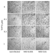
Viral cytopathic effects in Vero E6 cells after infection with SARS-CoV-2 and subsequent (single) PDI treatment. Three days after infection, cell cultures showed clear signs of viral cytopathic effects as a consequence of SARS-CoV-2 infection. When infection was followed by one single PDI for 10 min (light dose 7.8 J/cm2) with THPTS at indicated concentrations, viral cytopathic effects were mitigated in a concentration-dependent manner. At 1 µM THPTS, toxic effects of the PDI treatment were observed. Scale bar indicates 100 µm.
Figure 4
Figure 4
Viral cytopathic effects in Vero E6 cells after infection with SARS-CoV-2 and subsequent repeated PDI treatment. Three days after infection, cell cultures showed clear signs of viral cytopathic effects as a consequence of SARS-CoV-2 infection. When infection was followed by repeated PDI for 5 min every 4 h (light dose 3.9 J/cm2) with THPTS at indicated concentrations, viral cytopathic effects were mitigated in a concentration-dependent manner. At 1 µM THPTS, toxic effects of the PDI treatment were observed. Scale bar indicates 100 µm.
Figure 5
Figure 5
Results from photodynamic inactivation treatment of Vero cell cultures after infection with SARS-CoV-2. (A) Viral RNA was detected after SARS-CoV-2 infection of Vero cell cultures and subsequent single (5 min) or repeated PDI (5 min every 4 h for 3 d) at different photosensitizer (PS) concentrations. (B) Cell viability was determined by an MTT-based assay for mitochondrial activity. Data are presented as mean and SD. * p < 0.05 when compared to respective samples treated with 0 µM THPTS.

References

    1. World Health Organization WHO COVID-19 Dashboard. 2022. [(accessed on 16 May 2022)]. Available online: https://covid19.who.int.
    1. van Doremalen N., Bushmaker T., Morris D.H., Holbrook M.G., Gamble A., Williamson B.N., Tamin A., Harcourt J.L., Thornburg N.J., Gerber S.I., et al. Aerosol and Surface Stability of SARS-CoV-2 as Compared with SARS-CoV-1. N. Engl. J. Med. 2020;382:1564–1567. doi: 10.1056/NEJMc2004973. - DOI - PMC - PubMed
    1. Lodder W., de Roda Husman A.M. SARS-CoV-2 in Wastewater: Potential Health Risk, but Also Data Source. Lancet Gastroenterol. Hepatol. 2020;5:533–534. doi: 10.1016/S2468-1253(20)30087-X. - DOI - PMC - PubMed
    1. Abrahamse H., Hamblin M.R. New Photosensitizers for Photodynamic Therapy. Biochem. J. 2016;473:347–364. doi: 10.1042/BJ20150942. - DOI - PMC - PubMed
    1. Wainwright M. Anti-Infective Dyes in the Time of COVID. Dyes Pigm. 2021;196:109813. doi: 10.1016/j.dyepig.2021.109813. - DOI - PMC - PubMed

Publication types

LinkOut - more resources