. 2022 Jun 20;14(6):1344.
doi: 10.3390/v14061344.

The Viral Susceptibility of the Haloferax Species

Affiliations

The Viral Susceptibility of the Haloferax Species

Zaloa Aguirre Sourrouille et al. Viruses. .

Abstract

Viruses can infect members of all three domains of life. However, little is known about viruses infecting archaea and the mechanisms that determine their host interactions are poorly understood. Investigations of molecular mechanisms of viral infection rely on genetically accessible virus-host model systems. Euryarchaea belonging to the genus Haloferax are interesting models, as a reliable genetic system and versatile microscopy methods are available. However, only one virus infecting the Haloferax species is currently available. In this study, we tested ~100 haloarchaeal virus isolates for their infectivity on 14 Haloferax strains. From this, we identified 10 virus isolates in total capable of infecting Haloferax strains, which represented myovirus or siphovirus morphotypes. Surprisingly, the only susceptible strain of all 14 tested was Haloferax gibbonsii LR2-5, which serves as an auspicious host for all of these 10 viruses. By applying comparative genomics, we shed light on factors determining the host range of haloarchaeal viruses on Haloferax. We anticipate our study to be a starting point in the study of haloarchaeal virus-host interactions.

Keywords: Haloferax; Haloferax gibbonsii LR2-5; archaeal virus; haloarchaea; host range.


Conflict of interest statement

The authors declare no conflict of interest.

Figures
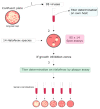
Figure 1
Figure 1
A schematic overview of the virus–host screen. Step 1: Fresh virus stocks made from confluent or semi-confluent plates were prepared on their own host strains and the titers were determined on their own host strains. Step 2: 95 virus stocks (undiluted and 10−2 dilution) were spotted on lawns of 14 Haloferax strains. MGM medium was used as a negative control (CTL). Step 3: After incubation at 37 °C, all virus-Haloferax pairs that resulted in growth inhibition on the spot-on lawn-assay were further tested by plaque assay by making serial dilutions of the virus stock and plating with the Haloferax strains to be tested. Viral plaques observed on Haloferax were counted, the titers were determined, and positive virus-Haloferax pairs were noted in Table 1.
Figure 2
Figure 2
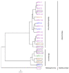
Phylogenetic tree of viruses belonging to the Haloferuviridae and Hafunaviridae families based on average nucleotide identity (ANI) values calculated using VIRIDIC software. Viruses infecting LR2-5 are surrounded by a box. Scale bar represents the number of substitutions per nucleotide position. Place of isolation: pink Senegal, purple Thailand, green Slovenia, orange Israel, and blue Italy.
Figure 3
Figure 3
(A) Isolation sites of the haloferacalesviruses, mincapviruses, and retbasiphovirus (see also Supplementary Table S2). Schemes of the viral morphologies that were observed in each group are also indicated (see also Table 1). LR2-5 infecting viruses are circled with a dash line. (B) Schematic representation of the myovirus and siphovirus virion morphologies (not in scale) and a typical genome organization consisting of different functional modules of tailed archaeal viruses.
Figure 4
Figure 4
Schematic genomic alignment of the (A) mincapviruses, and (B) haloferacalesviruses. Grey bars represent homologous genomic regions. The level of nucleotide identity is reflected by the intensity of grey. Genes encoding major capsid protein (orange) and adhesins (pink) are indicated. The LR2-5 infecting viruses are circled with dashed lines. Identical or very similar genomes are shown as one and virus names are separated by /. Figures are prepared with Easyfig.
Figure 5
Figure 5
Comparison of the adhesin and tail fiber gene sequence phylogenies to the ability of viruses to infect LR2-5. (a,c) Distribution of Markov jumps (95% HPD) for (i) real states (pink) and (ii) randomized states (grey). (b,d) Bayesian maximum clade credibility tree with discrete trait reconstruction based on adhesin or tail fiber gene sequences, respectively. Asterisk indicates node posterior probabilities higher than 0.8, and viruses in the boxes can infect LR2-5. Color code indicates the genus; blue Mincapvirus, brown Haloferacalesvirus.

References

    1. Borrel G., Brugère J.-F., Gribaldo S., Schmitz R.A., Moissl-Eichinger C. The Host-Associated Archaeome. Nat. Rev. Microbiol. 2020;18:622–636. doi: 10.1038/s41579-020-0407-y. - DOI - PubMed
    1. Lloyd K.G., May M.K., Kevorkian R.T., Steen A.D. Meta-Analysis of Quantification Methods Shows That Archaea and Bacteria Have Similar Abundances in the Subseafloor. Appl. Environ. Microbiol. 2013;79:7790–7799. doi: 10.1128/AEM.02090-13. - DOI - PMC - PubMed
    1. Forterre P., Prangishvili D. The Origin of Viruses. Res. Microbiol. 2009;160:466–472. doi: 10.1016/j.resmic.2009.07.008. - DOI - PubMed
    1. Hartman R., Munson-McGee J., Young M.J., Lawrence C.M. Survey of High-Resolution Archaeal Virus Structures. Curr. Opin. Virol. 2019;36:74–83. doi: 10.1016/j.coviro.2019.05.008. - DOI - PubMed
    1. Baquero D.P., Liu Y., Wang F., Egelman E.H., Prangishvili D., Krupovic M. Structure and Assembly of Archaeal Viruses. 1st ed. Volume 108. Elsevier Inc.; Amsterdam, The Netherlands: 2020. - PubMed

Publication types

LinkOut - more resources