. 2022 Sep 7;30(9):3066-3077.
doi: 10.1016/j.ymthe.2022.06.013. Epub 2022 Jun 22.

Eliciting anti-cancer immunity by genetically engineered multifunctional exosomes

Affiliations

Eliciting anti-cancer immunity by genetically engineered multifunctional exosomes

Qinqin Cheng et al. Mol Ther. .

Abstract

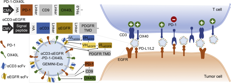
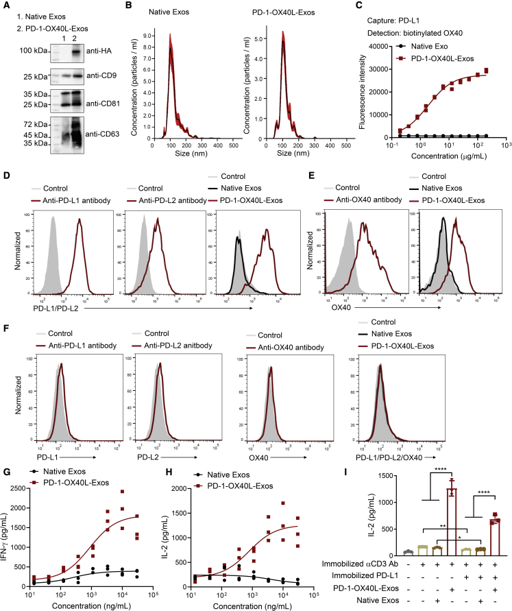
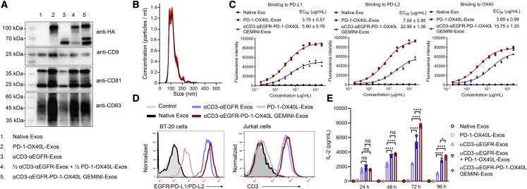
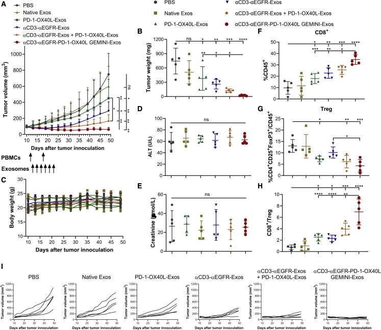
Exosomes are cell-derived nanovesicles involved in regulating intercellular communications. In contrast to conventional nanomedicines, exosomes are characterized by unique advantages for therapeutic development. Despite their major successes in drug delivery, the full potential of exosomes for immunotherapy remains untapped. Herein we designed genetically engineered exosomes featured with surfaced-displayed antibody targeting groups and immunomodulatory proteins. Through genetic fusions with exosomal membrane proteins, Expi293F cell-derived exosomes were armed with monoclonal antibodies specific for human T-cell CD3 and epidermal growth factor receptor (EGFR) as well as immune checkpoint modulators, programmed death 1 (PD-1) and OX40 ligand (OX40L). The resulting genetically engineered multifunctional immune-modulating exosomes (GEMINI-Exos) can not only redirect and activate T cells toward killing EGFR-positive triple negative breast cancer (TNBC) cells but also elicit robust anti-cancer immunity, giving rise to highly potent inhibition against established TNBC tumors in mice. GEMINI-Exos represent candidate agents for immunotherapy and may offer a general strategy for generating exosome-based immunotherapeutics with desired functions and properties.

Keywords: exosomes; extracellular vesicles; immunotherapy; protein engineering; synthetic biology; triple negative breast cancer.


Conflict of interest statement

Declaration of interests The authors declare no competing interests.

Figures

None
Graphical abstract
Figure 1
Figure 1
Schematic of αCD3-αEGFR-PD-1-OX40L GEMINI-Exos for targeted cancer immunotherapy HA, hemagglutinin; PD-1, programmed death 1; OX40L, OX40 ligand; αCD3, anti-CD3; αEGFR, anti-EGFR; scFv, single-chain variable fragment; PDGFR TMD, transmembrane domain of human platelet-derived growth factor receptor; PD-L1/L2, programmed death-ligand 1/ligand 2.
Figure 2
Figure 2
Generation and characterization of PD-1-OX40L-Exos (A) Immunoblot analysis of purified exosomes. (B) Size distribution of native exosomes and PD-1-OX40L-Exos. (C) Sandwich ELISA analysis of the binding of PD-1-OX40L-Exos to human PD-L1 and OX40. Recombinant human PD-L1 and biotinylated OX40 were used as capture and detection reagents, respectively. Data are shown as mean ± SD of duplicates. (D–F) Flow cytometry of the binding of PD-1-OX40L-Exos to IFN-γ stimulated BT-20 cells (D), activated human T cells (E), and MDA-MB-468 cells (F). Anti-PD-L1, anti-PD-L2, and anti-OX40 antibodies were used as positive controls. Arrows show signal levels of indicated target proteins being detected after the incubation with antibodies, native Exos, or PD-1-OX40L-Exos. (G and H) Dose-dependent activation of human T cells by PD-1-OX40L-Exos. Human PBMCs were incubated with pre-coated anti-human CD3 monoclonal antibody in the presence of various concentrations of PD-1-OX40L-Exos or native exosomes for 48 h. The levels of secreted IFN-γ (G) and IL-2 (H) were measured by ELISA. Data are shown as mean ± SD of triplicates. (I) PD-1-OX40L-Exos restore T-cell activation from PD-L1-mediated inhibition. Human PBMCs were incubated with pre-coated anti-human CD3 monoclonal antibody without or with pre-coated human PD-L1 in the absence or presence of 10 μg mL−1 PD-1-OX40L-Exos or native exosomes for 48 h. The levels of secreted IL-2 were measured by ELISA. Data are shown as mean ± SD of triplicates. ∗p < 0.05 and ∗∗∗∗p < 0.0001 (two-tailed unpaired t test).
Figure 3
Figure 3
Generation and characterization of αCD3-αEGFR-PD-1-OX40L GEMINI-Exos (A) Immunoblot analysis of purified exosomes. (B) Size distribution of αCD3-αEGFR-PD-1-OX40L GEMINI-Exos. (C) ELISA analysis of the binding of αCD3-αEGFR-PD-1-OX40L GEMINI-Exos to human PD-L1, PD-L2, and OX40. PD-1-OX40L-Exos, αCD3-αEGFR-PD-1-OX40L GEMINI-Exos, and native exosomes at various concentrations were coated on 96-well ELISA plates overnight, followed by incubation with recombinant PD-L1-Fc, PD-L2-Fc, or OX40-Fc and detection with an anti-human IgG-HRP. Data are shown as mean ± SD of duplicates. (D) Flow cytometry of the binding of αCD3-αEGFR-PD-1-OX40L GEMINI-Exos to BT-20 cells (EGFR+ PD-L1+) and Jurkat cells (CD3+). Arrows show signal levels of indicated target proteins being detected after the incubation with various types of exosomes. (E) Time-dependent activation of human T cells by αCD3-αEGFR-PD-1-OX40L GEMINI-Exos. Human PBMCs were incubated with BT-20 cells at a ratio of 2:1 for 24 to 96 h in the presence of native exosomes, PD-1-OX40L-Exos, αCD3-αEGFR-Exos, a mixture (1:1) of PD-1-OX40L- and αCD3-αEGFR-Exos, or αCD3-αEGFR-PD-1-OX40L GEMINI-Exos. The levels of secreted IL-2 were measured by ELISA. Data are shown as mean ± SD of triplicates. ns = not significant, ∗p < 0.05, and ∗∗∗∗p < 0.0001 (ordinary one-way ANOVA test).
Figure 4
Figure 4
In vivo evaluation of αCD3-αEGFR-PD-1-OX40L GEMINI-Exos (A) Anti-tumor activity of GEMINI-Exos. BT-20 cells were subcutaneously implanted into the flank of female NSG mice (n = 5). In vitro expanded human PBMCs from the same healthy donor were intraperitoneally injected into mice on days 12 and 18 post tumor implantation. One day post the first PBMC administration, mice were treated with PBS or different types of exosomes (10 mg/kg for monotherapy and 20 mg/kg for combination therapy) every other day for a total of six times via intravenous injections. Data are shown as mean ± SD (n = 5). ns = not significant, ∗p < 0.05, and ∗∗p < 0.01(one-way repeated measures ANOVA test with the Geisser-Greenhouse correction). (B) Tumor weights at the end of study. (C) Body weights of mice during the study. (D) ALT activities in plasma at the end of study. (E) Creatinine concentrations in plasma at the end of study. (F) Percentage CD8+ T cells in CD45+ cells in tumors. (G) Percentages of CD4+ CD25+ FoxP3+ Tregs in CD45+ cells in tumors. (H) CD8+ T cell/Treg ratios in tumors. At the end of the study, tumors were harvested and disaggregated into single-cell suspensions. After immunostaining, cells were analyzed by flow cytometry for the expression of CD45, CD4, CD8, CD25, and FoxP3. (I) Tumor growth curves for individual mice during the study. Data in (B, D–H) are shown as mean ± SD (n = 5). ns = not significant, ∗p < 0.05, ∗∗p < 0.01, ∗∗∗p < 0.001, and ∗∗∗∗p < 0.0001 (ordinary one-way ANOVA test).

References

    1. Doyle L.M., Wang M.Z. Overview of extracellular vesicles, their origin, composition, purpose, and methods for exosome isolation and analysis. Cells. 2019;8:727. doi: 10.3390/cells8070727. - DOI - PMC - PubMed
    1. Kalluri R., LeBleu V.S. The biology, function, and biomedical applications of exosomes. Science. 2020;367:eaau6977. doi: 10.1126/science.aau6977. - DOI - PMC - PubMed
    1. Ståhl A.L., Johansson K., Mossberg M., Kahn R., Karpman D. Exosomes and microvesicles in normal physiology, pathophysiology, and renal diseases. Pediatr. Nephrol. 2019;34:11–30. - PMC - PubMed
    1. Boulanger C.M., Loyer X., Rautou P.-E., Amabile N. Extracellular vesicles in coronary artery disease. Nat. Rev. Cardiol. 2017;14:259–272. - PubMed
    1. Maia J., Caja S., Strano Moraes M.C., Couto N., Costa-Silva B. Exosome-based cell-cell communication in the tumor microenvironment. Front. Cell Dev. Biol. 2018;6:18. doi: 10.3389/fcell.2018.00018. - DOI - PMC - PubMed

Publication types

MeSH terms

Substances