

A Novel Technique for the Evaluation and Interpretation of Elastography in Salivary Gland Involvement in Primary Sjögren Syndrome
- PMID: 35746947
- PMCID: PMC9210135
- DOI: 10.3389/fmed.2022.913589
A Novel Technique for the Evaluation and Interpretation of Elastography in Salivary Gland Involvement in Primary Sjögren Syndrome
Abstract
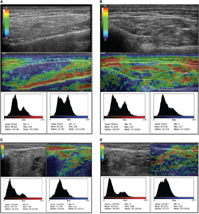
Ultrasound (US) of major salivary glands (MSG) evaluates echogenicity, border features and vascularization, with elastography, it can detect tissue elasticity and glandular fibrosis, related to inflammation in Primary Sjögren's syndrome (pSS). This study aimed to develop a novel technique by pixel analysis for evaluation and interpretation of elastography in MSG in pSS. A cross-sectional and observational multicenter study was conducted. The US of MSG performed in orthogonal planes in grayscale, Doppler, and shear-wave elastography. For elastography images of each gland were analyzed with the open-source program ImageJ to perform a pixel analysis. Statistical analysis was performed with the IBM-SPSS v25 program. Fifty-nine women with a mean age of 57.69 (23-83) years were recruited; pSS mean duration of 87 (5-275) months, and 12 healthy women without sicca symptoms as a control group with a mean age of 50.67 (42-60) years. Intragroup analysis showed p-values >0.05 between sicca symptoms, ocular/dryness tests, biopsy, US, and pixel analysis; correlation between Hocevar and pixel analysis was not found (rho < 0.1, p >0.5). MSG anatomical size was 41.7 ± 28.2 mm vs. 67.6 ± 8.8 mm (p ≤ 0.0001); unstimulated whole saliva flow rate was 0.80 ± 0.80 ml/5 min vs. 1.85 ± 1.27 ml/5 min (p = 0.016). The elastography values (absolute number of pixels) were 572.38 ± 99.21 vs. 539.69 ± 93.12 (p = 0.290). A cut-off point risk for pSS identified with less than 54% of red pixels in the global MSG mass [OR of 3.8 95% CI (1.01-15.00)]. Pixel analysis is a new tool that could lead to a better understanding of the MSG chronic inflammatory process in pSS.
Keywords: elastography; major salivary glands; pixel analysis; primary Sjögren syndrome; ultrasound.
Copyright © 2022 Barbosa-Cobos, Torres-González, Meza-Sánchez, Ventura-Ríos, Concha-Del-Río, Ramírez-Bello, Álvarez-Hernández, Meléndez-Mercado, Enríquez-Sosa, Samuria-Flores, Lugo-Zamudio and Hernández-Díaz.
Conflict of interest statement
The authors declare that the research was conducted in the absence of any commercial or financial relationships that could be construed as a potential conflict of interest.
Figures
References
-
- Rischmueller M, Spurrier A. Can Sjögren’s syndrome diagnosis and evaluation be stretched by elastography? Int J Rheum Dis. (2019) 22:172–4. - PubMed
-
- Shiboski CH, Shiboski SC, Seror R, Criswell LA, Labetoulle M, Lietman TM, et al. 2016 American college of rheumatology/European league against rheumatism classification criteria for primary Sjogren’s syndrome: a consensus and data-driven methodology involving three international patient cohorts. Arthritis Rheumatol. (2017) 69:35–45. - PMC - PubMed
-
- Silvers AR, Som PM. Salivary glands. Radiol Clin North Am. (1998) 36:941–66. - PubMed
-
- Kroese FGM, Haacke EA, Bombardieri M. The role of salivary gland histopathology in primary Sjögren’s syndrome promises and pitfalls. Clin Exp Rheumatol. (2018) 36(Suppl. 112):222–33. - PubMed
LinkOut - more resources
Full Text Sources