Upregulation of osteoprotegerin inhibits tert-butyl hydroperoxide-induced apoptosis of human chondrocytes
- PMID: 35747145
- PMCID: PMC9204554
- DOI: 10.3892/etm.2022.11397
Upregulation of osteoprotegerin inhibits tert-butyl hydroperoxide-induced apoptosis of human chondrocytes
Abstract
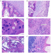
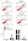
Necrosis of the femoral head (NFH) is an orthopedic disease characterized by a severe lack of blood supply to the femoral head and a marked increase in intraosseous pressure. NFH is associated with numerous factors, such as alcohol consumption and hormone levels. The present study focused on the expression levels of osteoprotegerin (OPG) in NFH and the effect of OPG overexpression on chondrocyte apoptosis. The results demonstrated that OPG expression was markedly decreased in the femoral head of patients with NFH compared with normal femoral heads. Lentivirus-mediated overexpression of OPG in human chondrocytes reversed the decrease in cell viability and the increase in reactive oxygen species production induced by an oxidative stress-inducing factor, tert-butyl hydroperoxide. Flow cytometry and TUNEL assays revealed that OPG overexpression inhibited the apoptosis of chondrocytes. In addition, it was revealed that OPG exerted its anti-apoptotic effect mainly by promoting Bcl-2 expression and Akt phosphorylation and inhibiting caspase-3 cleavage and Bax expression. The present study revealed that OPG may be an important regulator of NFH.
Keywords: apoptosis; chondrocytes; necrosis of the femoral head; osteoprotegerin; tert-butyl hydroperoxide.
Copyright: © Ren et al.
Conflict of interest statement
The authors declare that they have no competing interests.
Figures


Similar articles
-
Effects of Wenyangbushen formula on the expression of VEGF, OPG, RANK and RANKL in rabbits with steroid-induced femoral head avascular necrosis.Mol Med Rep. 2015 Dec;12(6):8155-61. doi: 10.3892/mmr.2015.4478. Epub 2015 Oct 23. Mol Med Rep. 2015. PMID: 26496816
-
Osteoprotegerin deficiency leads to deformation of the articular cartilage in femoral head.J Mol Histol. 2016 Oct;47(5):475-83. doi: 10.1007/s10735-016-9689-9. Epub 2016 Aug 19. J Mol Histol. 2016. PMID: 27541035
-
Osteoprotegerin causes apoptosis of endothelial progenitor cells by induction of oxidative stress.Arthritis Rheum. 2013 Aug;65(8):2172-82. doi: 10.1002/art.37997. Arthritis Rheum. 2013. PMID: 23666878
-
Osteoprotegerin in breast cancer: beyond bone remodeling.Mol Cancer. 2015 Jun 10;14:117. doi: 10.1186/s12943-015-0390-5. Mol Cancer. 2015. PMID: 26054853 Free PMC article. Review.
-
Osteoprotegerin rich tumor microenvironment: implications in breast cancer.Oncotarget. 2016 Jul 5;7(27):42777-42791. doi: 10.18632/oncotarget.8658. Oncotarget. 2016. PMID: 27072583 Free PMC article. Review.
Cited by
-
Non-Alcoholic Fatty Liver Disease and Bone Tissue Metabolism: Current Findings and Future Perspectives.Int J Mol Sci. 2023 May 8;24(9):8445. doi: 10.3390/ijms24098445. Int J Mol Sci. 2023. PMID: 37176153 Free PMC article. Review.
References
LinkOut - more resources
Full Text Sources
Research Materials
