Sequential Increase in Complement Factor I, iC3b, and Cells Expressing CD11b or CD14 in Cutaneous Vasculitis
- PMID: 35747245
- PMCID: PMC9213176
- DOI: 10.1155/2022/3888734
Sequential Increase in Complement Factor I, iC3b, and Cells Expressing CD11b or CD14 in Cutaneous Vasculitis
Abstract
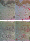
Mast cells contribute to the pathogenesis of cutaneous vasculitis through complement C3 that is cleaved to C3b and then to iC3b by complement factor I. The receptor of iC3b, CD11b, is expressed on neutrophils and monocytes and CD14 on monocytes. Their role in vasculitis is obscure. In this study, frozen skin biopsies from the nonlesional skin, initial petechial lesion, and palpable purpura lesion from 10 patients with immunocomplex-mediated small vessel vasculitis were studied immunohistochemically for complement factor I, iC3b, CD11b, and CD14. Peripheral blood mononuclear cells from 5 healthy subjects were used to study cell migration and cytokine secretion. Already, the nonlesional skin revealed marked immunostaining of complement factor I, iC3b, CD11b, and CD14, and their expression increased sequentially in initial petechial and palpable purpura lesions. Mast cell C3c correlated to iC3b, and both of them correlated to CD11b+ and CD14+ cells, in the nonlesional skin. The stimulation of mononuclear cells with 0.01-0.1 μg/ml iC3b induced cell migration in the transwell assay. C3a stimulated slightly interleukin-8 secretion, whereas 1 μg/ml iC3b inhibited it slightly, in 4/5 subjects. In conclusion, the C3-C3b-iC3b axis is activated already in the early vasculitis lesion leading to progressive accumulation of CD11b+ and CD14+ cells.
Copyright © 2022 Dina Rahkola et al.
Conflict of interest statement
The authors declare that they have no conflicts of interest.
Figures


References
-
- Vinen C. S. S., Turner D. R. R., Oliveira D. B. G. B. A central role for the mast cell in early phase vasculitis in the Brown Norway rat model of vasculitis: a histological study. International Journal of Experimental Pathology . 2004;85(3):165–174. doi: 10.1111/j.0959-9673.2004.00382.x. - DOI - PMC - PubMed
-
- Watanabe N., Akikusa B., Park S. Y., et al. Mast cells induce autoantibody-mediated vasculitis syndrome through tumor necrosis factor production upon triggering Fcgamma receptors. Blood, The Journal of the American Society of Hematology . 1999;94:3855–3863. - PubMed
MeSH terms
Substances
LinkOut - more resources
Full Text Sources
Medical
Research Materials
Miscellaneous

